Head & Neck
Κεφαλή & Τράχηλος
40 images

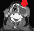
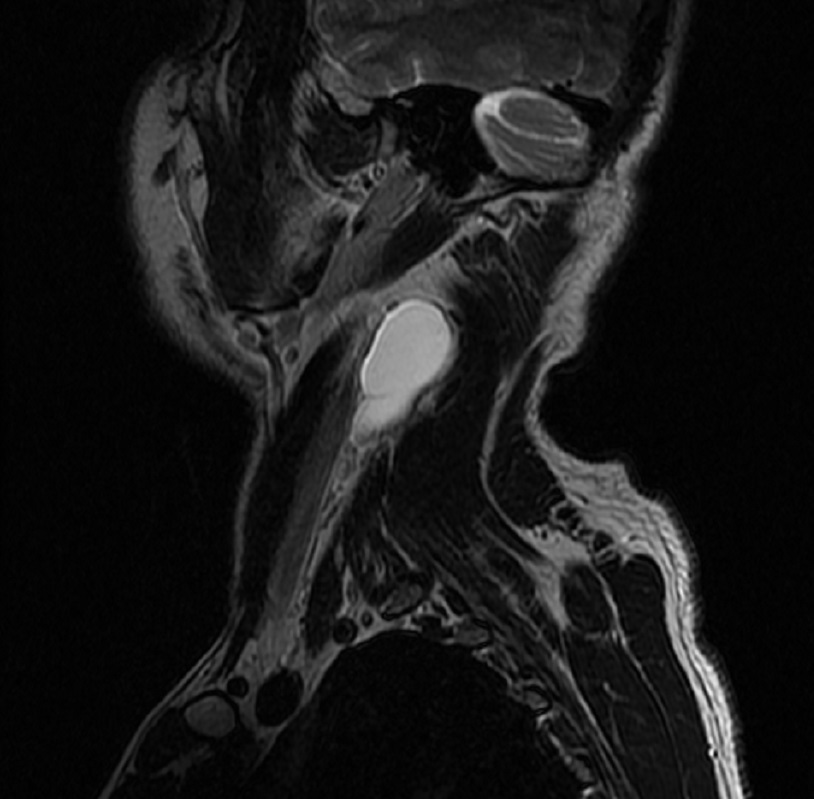
Neck CT Scan.Cystic lesion in close proximity with the hyoid bone.(Courtesy Dr.V.Penopoulos).
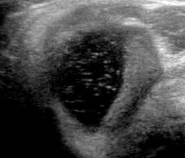
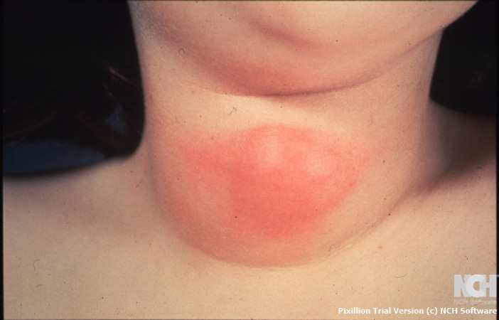
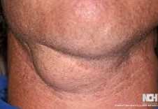
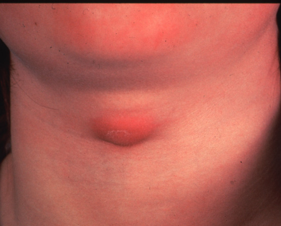
Neck U/S.Sizeable cystic lesion. Thyroglossal duct cyst carcinoma .(Courtesy Dr.V.Penopoulos).
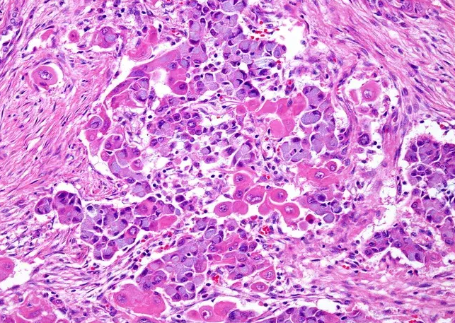
Mucinous cells with blue cytoplasm and flattened nuclei, intermixed with squamous cells containing dense eosinophilic cytoplasm and round nuclei. Courtesy Dr. V. Penopoulos.
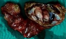
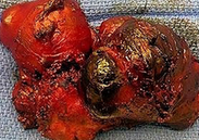
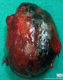
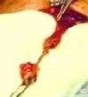
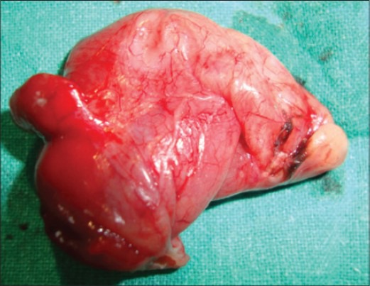
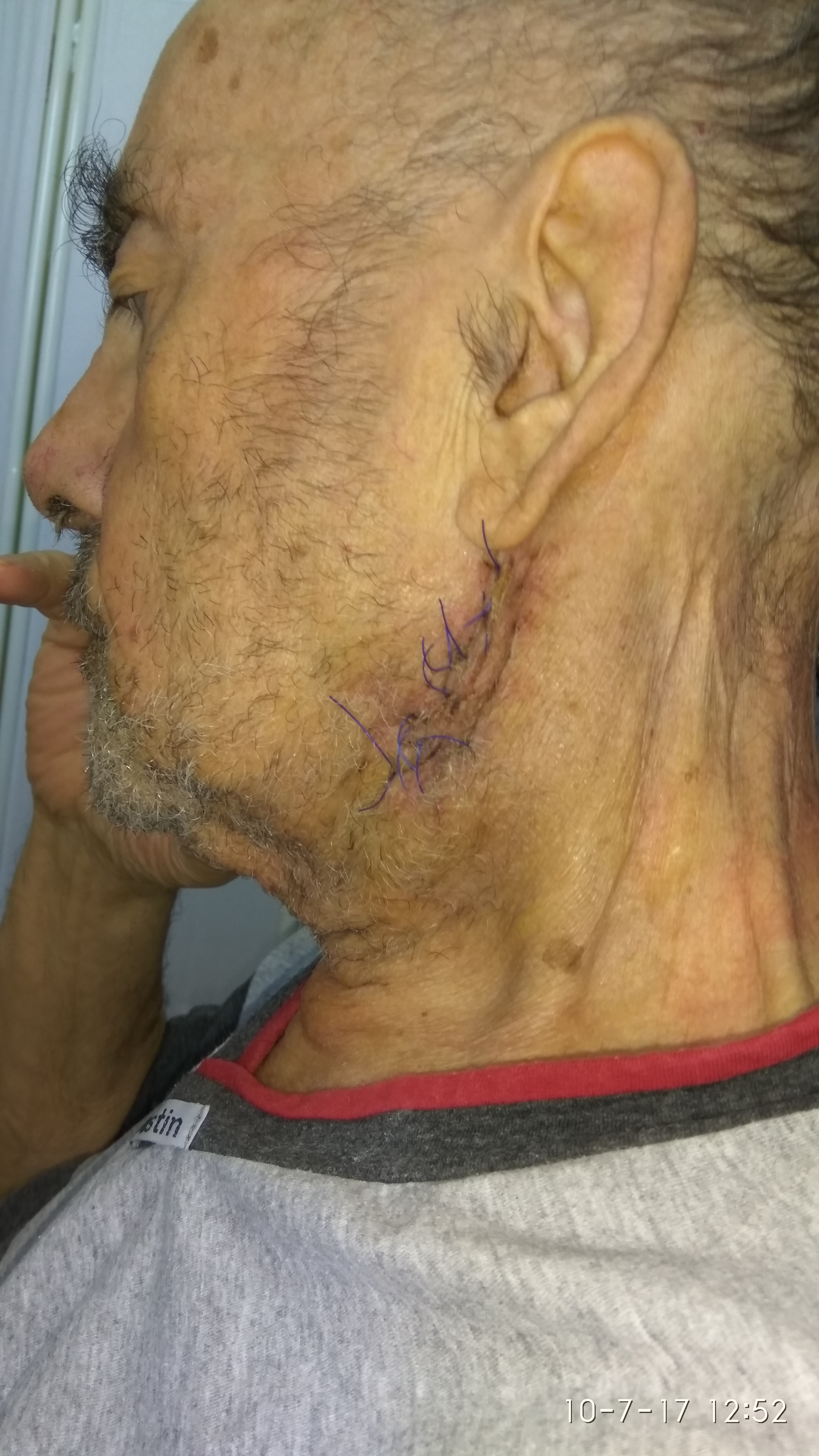
Specimen of left thyroid lobectomy and cystectomy.(Courtesy Dr.V.Penopoulos).
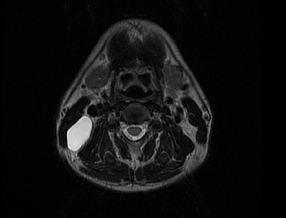
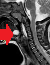
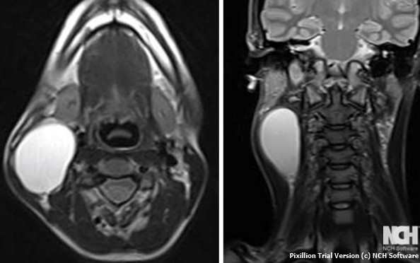
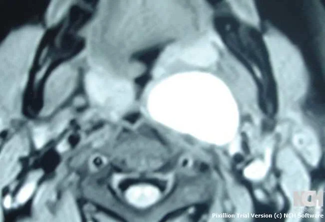
MRI images in the right neck.There is no significant solid or nodular component or surrounding infiltration.(Courtesy Dr.V.Penopoulos )
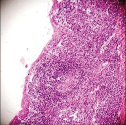
The branchial cleft cyst was lined by stratified squamous epithelium.Keratinaceous debris is noted in the lumen. Lymphoid aggregates with germinal centers are identified subjacent to the epithelium
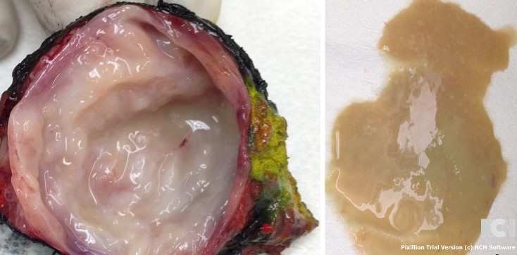
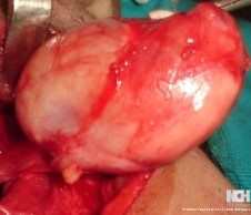
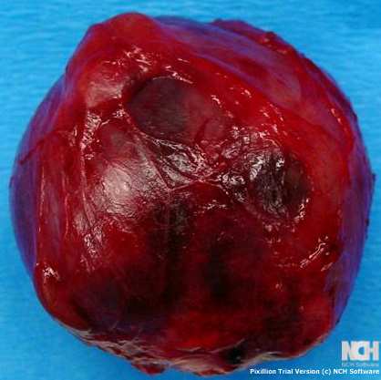
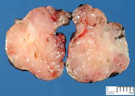
Gross examination of the excised cyst reveals a smooth surface lining of uniform thickness without papillary projections (Courtesy Dr. V. Penopoulos)
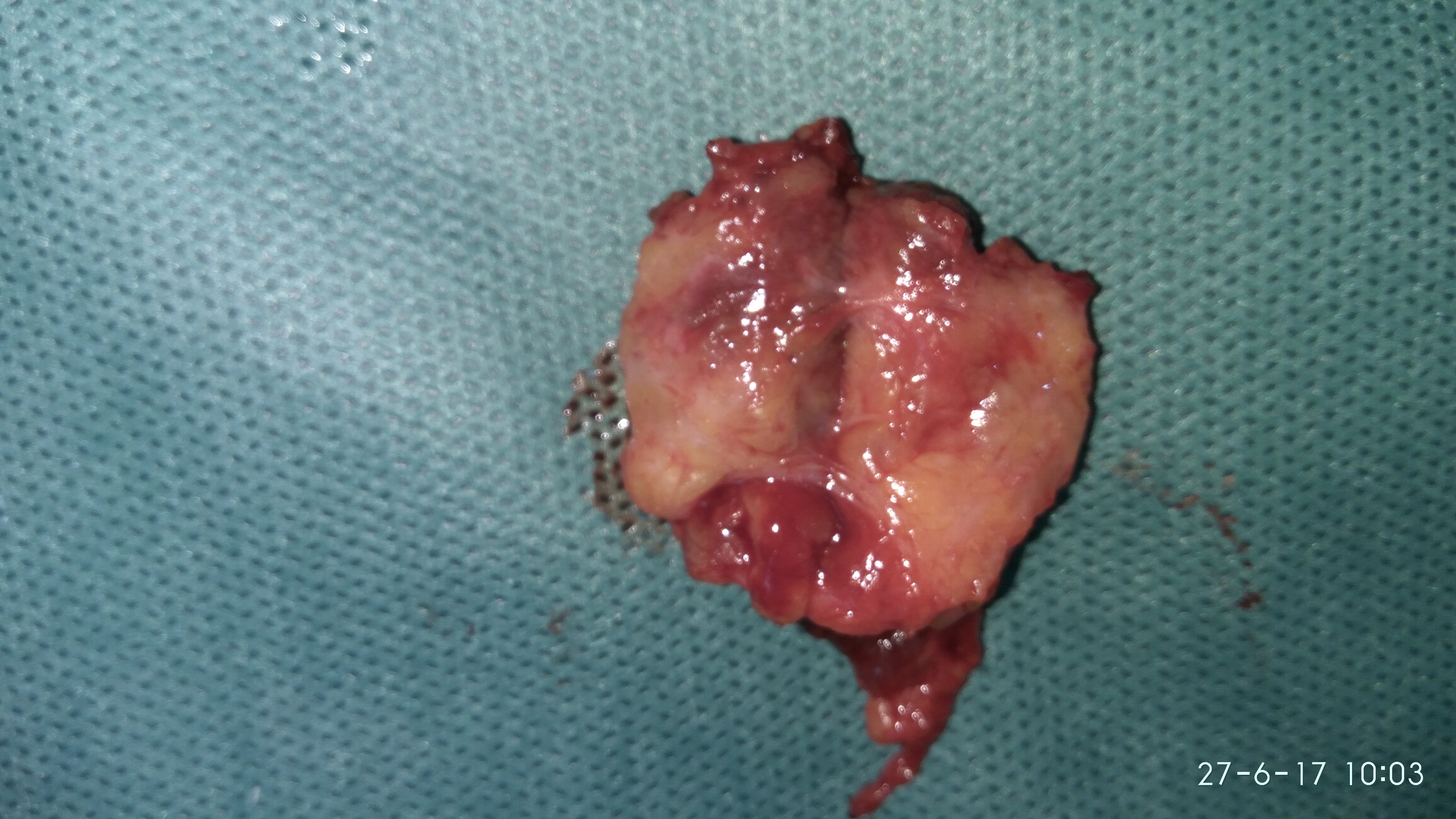

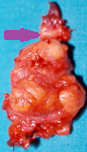
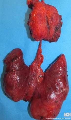
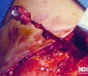
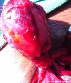
Surgical specimen of complete excision of thyreoglossal duct cyst.Purple arrow-Hyoid bone .Yellow arrow-Thyroglossal cyst.(Courtesy Dr.V.Penopoulos).
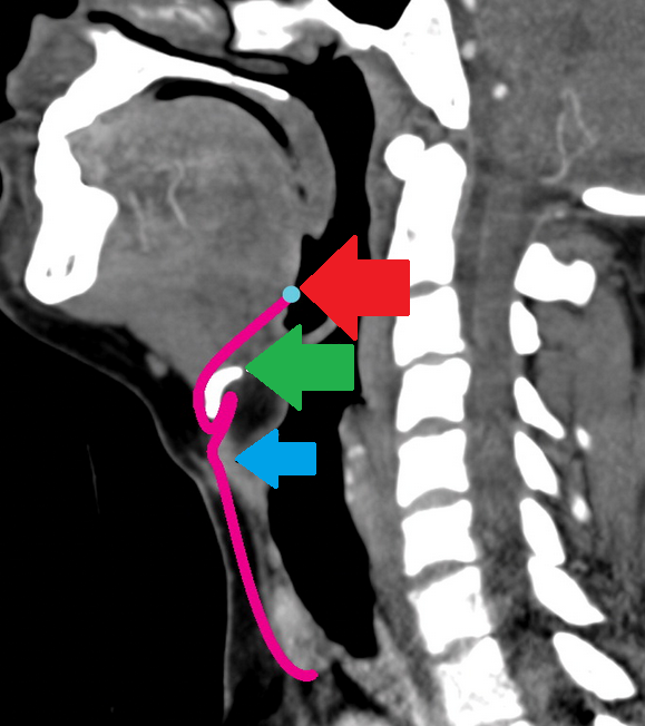
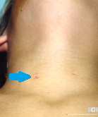
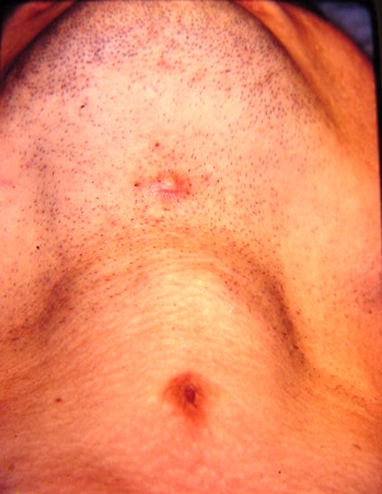
Red arrow-Foramen cecum.Green arrow-Hyoid bone.Light blue arrow-Thyroid cartilage.(Courtesy Dr.V.Penopoulos).
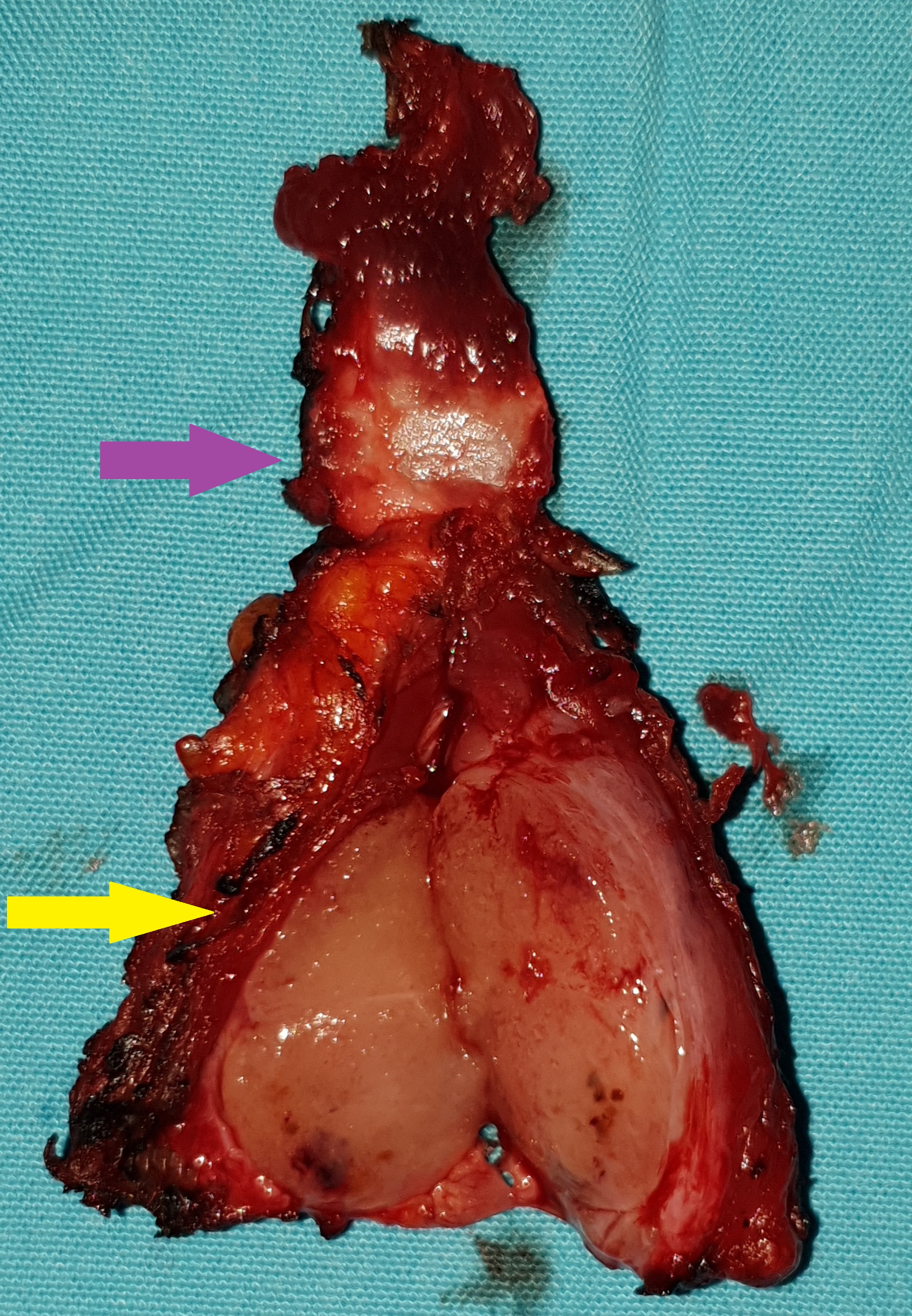
Surgical specimen of complete excision of thyreoglossal duct cyst.Purple arrow-Hyoid bone.Yellow arrow-Thyroglossal cyst.(Courtesy Dr.V.Penopoulos).