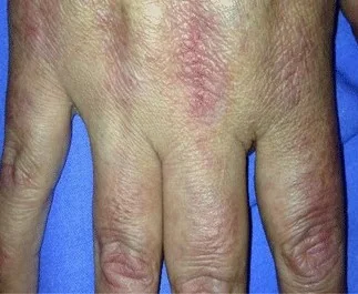
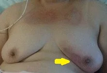
Breast
Μαστός
223 images


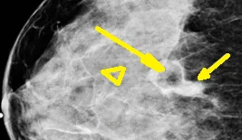
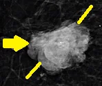
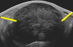
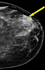
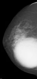
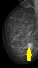


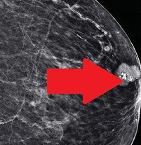


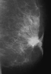
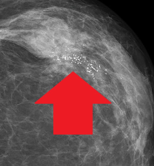
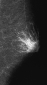
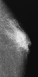

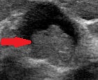


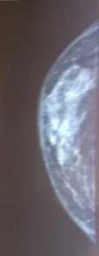


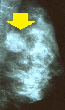
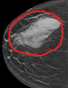
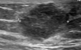
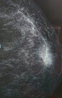
Digital Mammography.High density irregular mass right breast.(Courtesy Dr.V.Penopoulos).
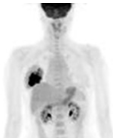
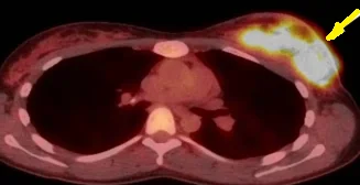
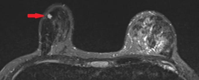
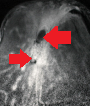
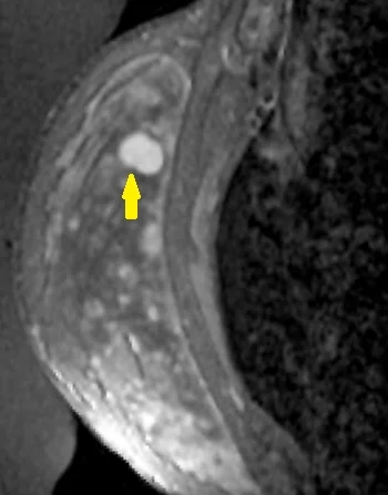
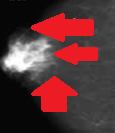
PET Scan. The hypermetabolic focus in the right breast is evident (Courtesy Dr. V. Penopoulos)
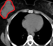
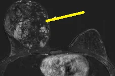
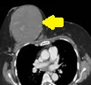
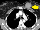
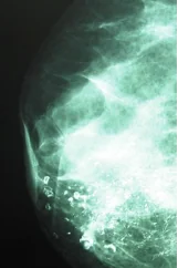
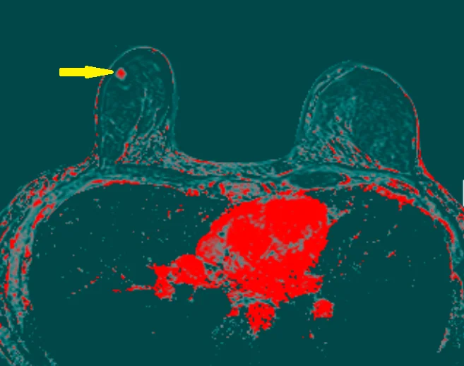
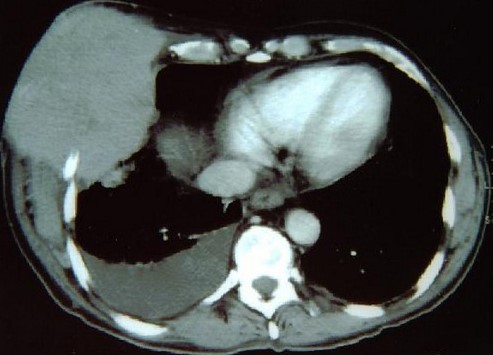
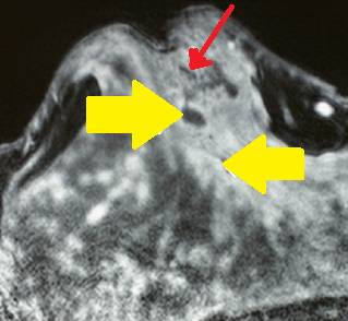
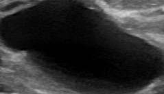
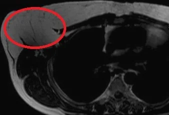
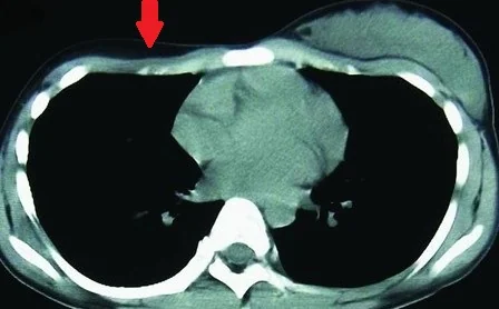
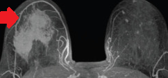
Chest CT Scan.Presence of a sizable, irregular shape mass in the right breast.(Courtesy Dr.V.Penopoulos).
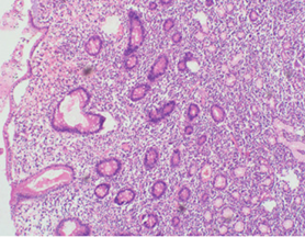
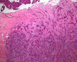
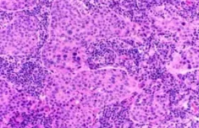
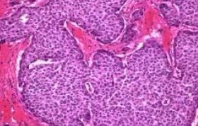
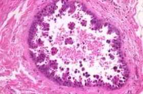
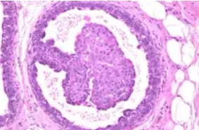
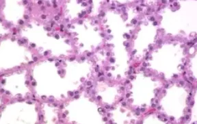
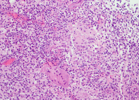
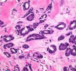
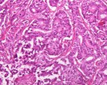
Presence of variably sized “crowded” follicles, composed of centrocytes, centroblasts, and infiltrates of lymphoid cells, along with lymphoepithelial lesions.(Courtesy Dr.V.Penopoulos).
Presence of variably sized “crowded” follicles, composed of centrocytes, centroblasts, and infiltrates of lymphoid cells, along with lymphoepithelial lesions.(Courtesy Dr.V.Penopoulos).
PET Scan. The hypermetabolic focus in the right breast is evident (Courtesy Dr. V. Penopoulos)
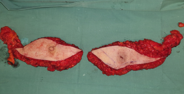
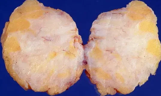
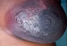
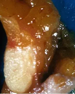
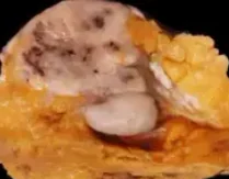
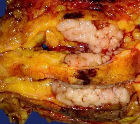
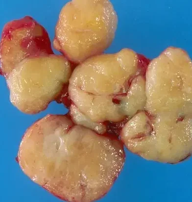
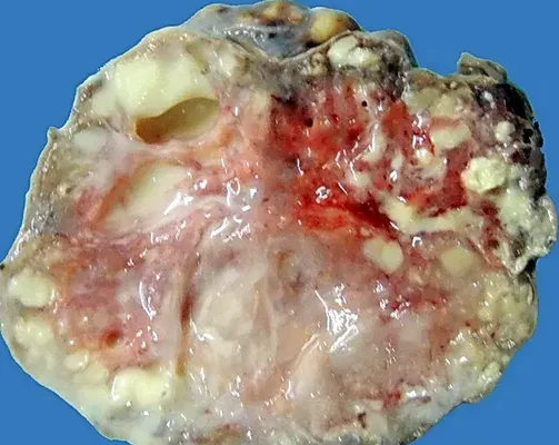
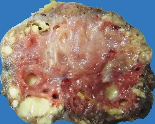
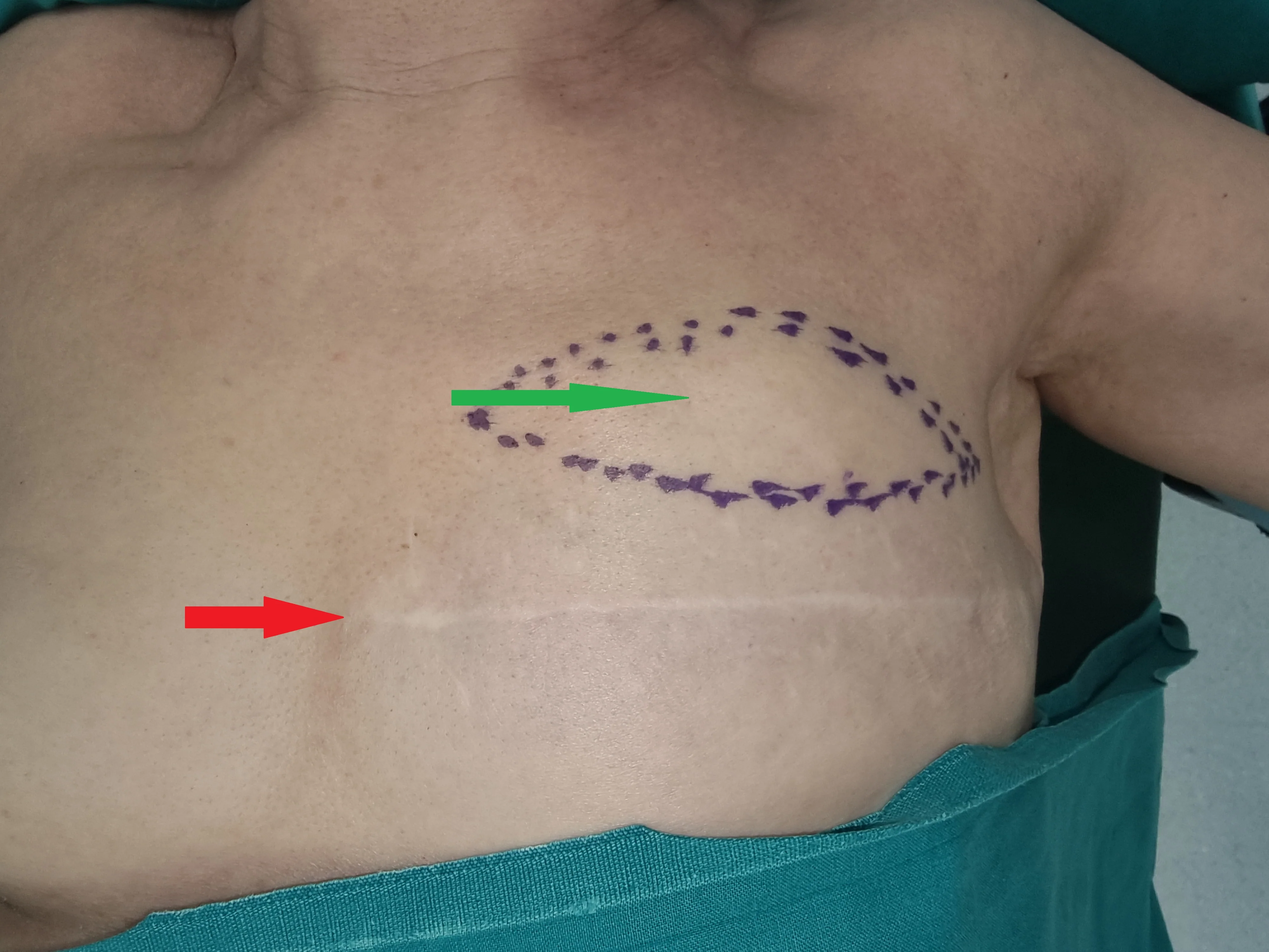
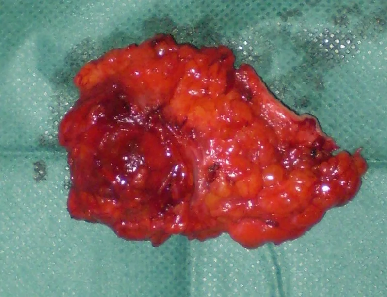
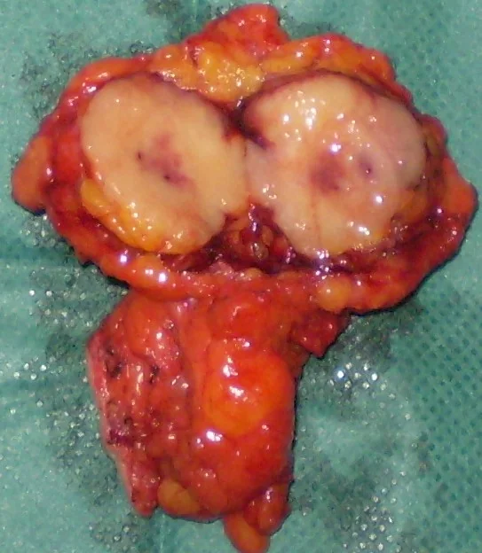
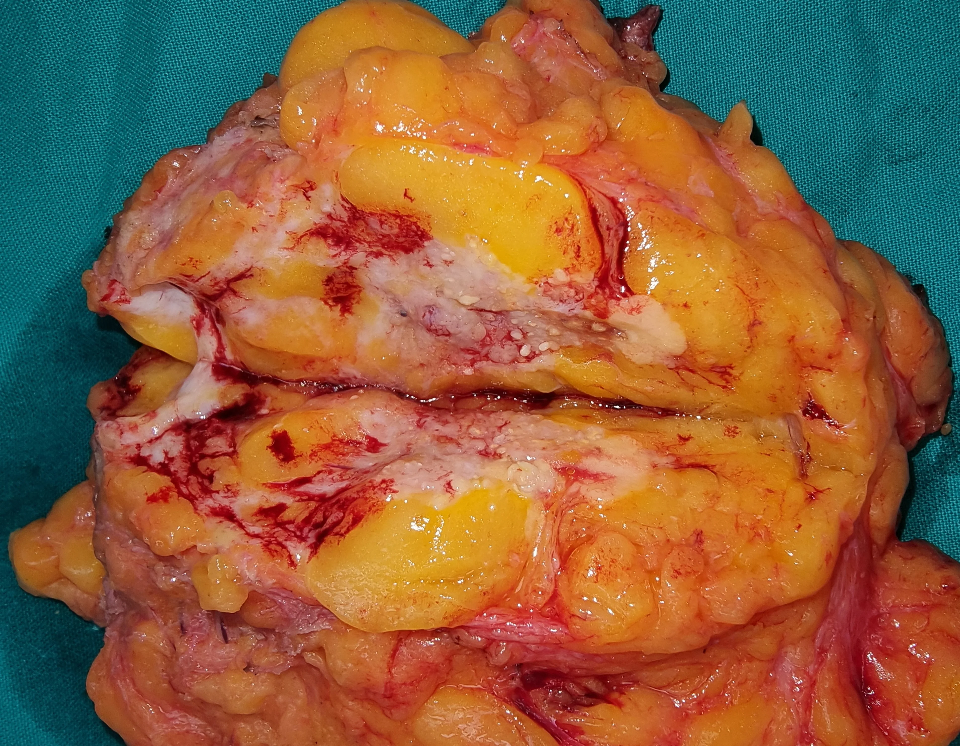
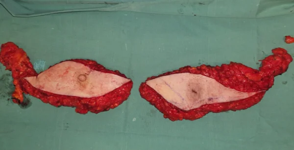
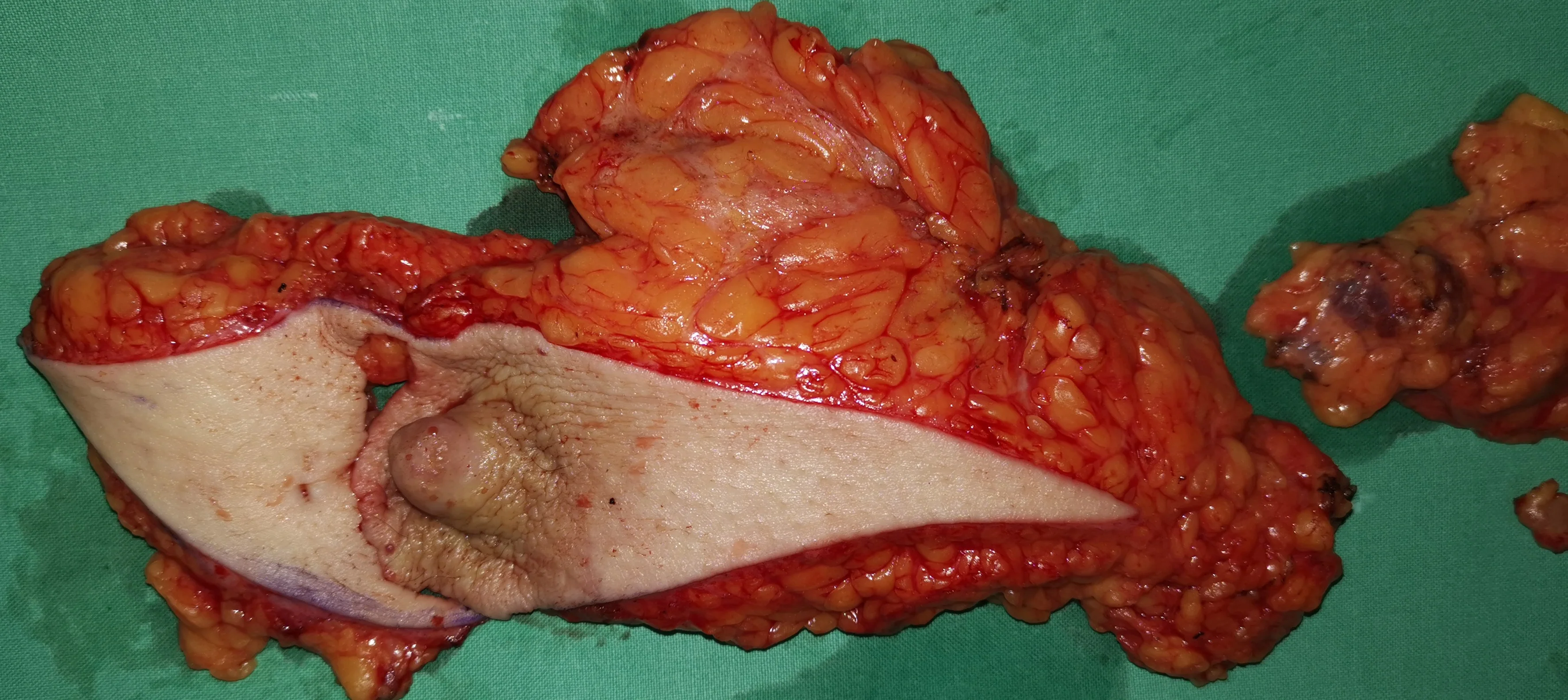
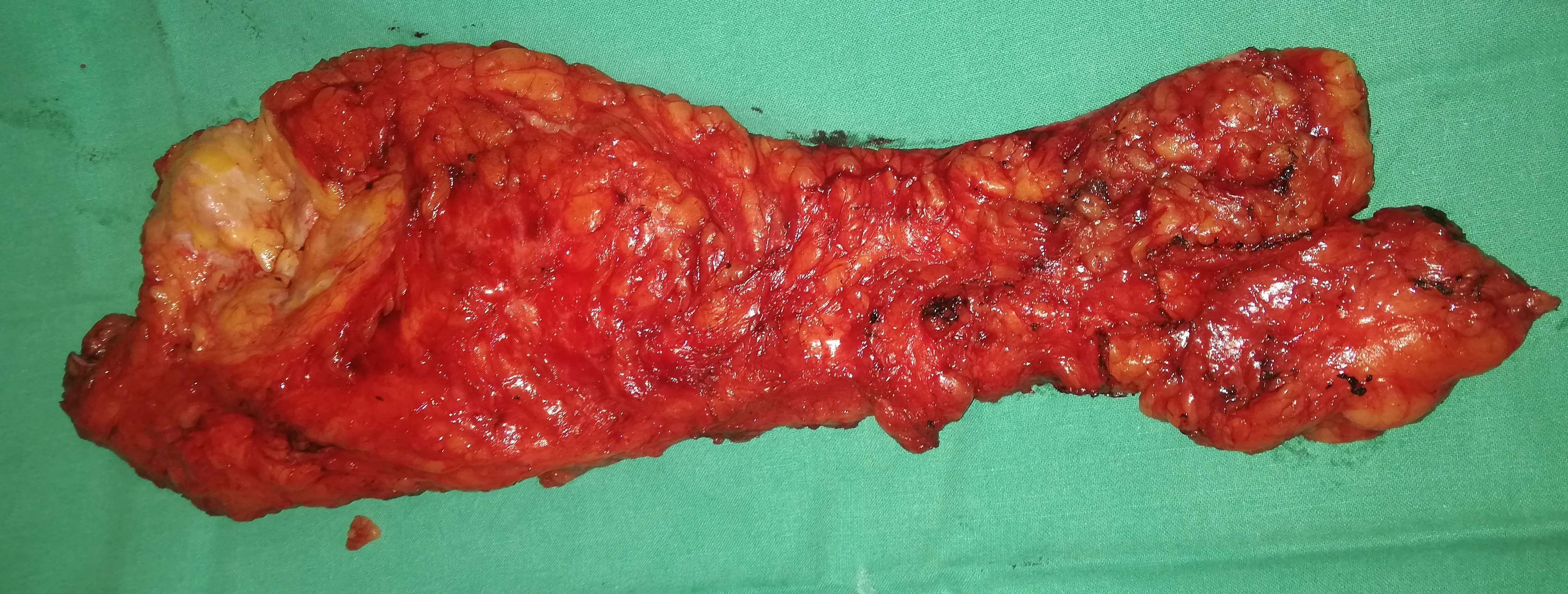
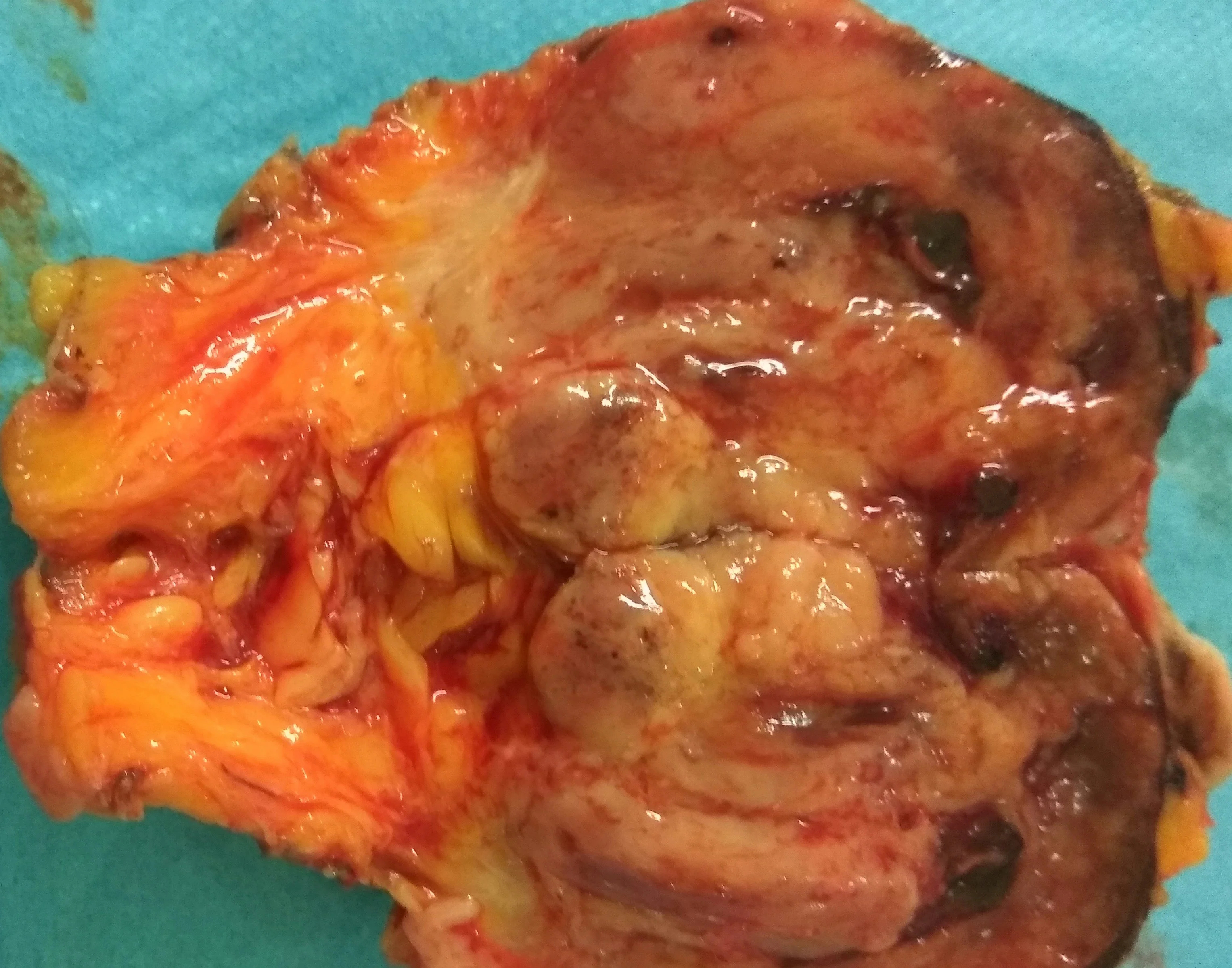
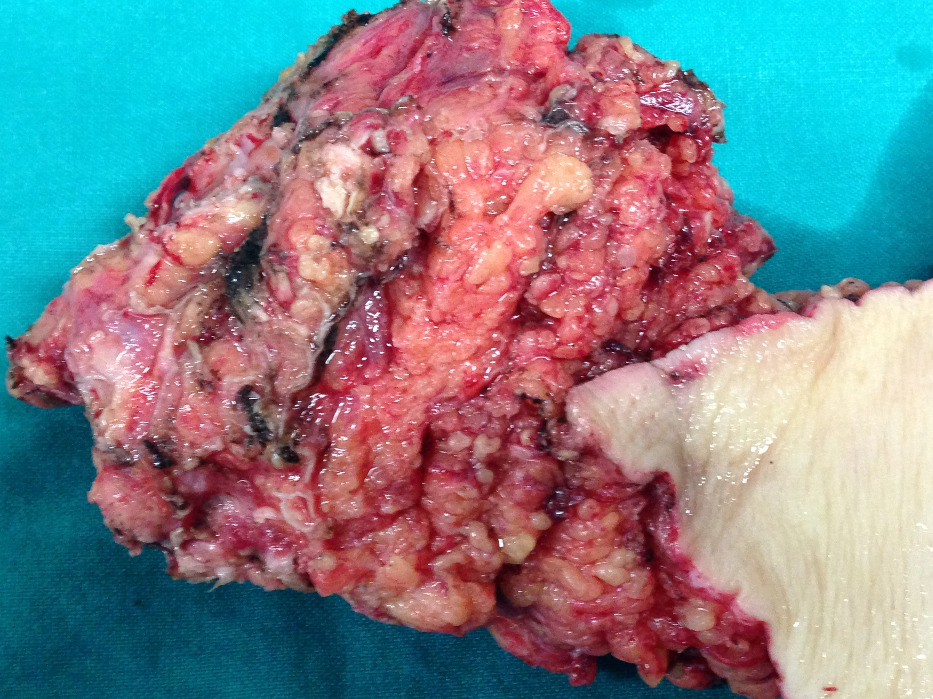
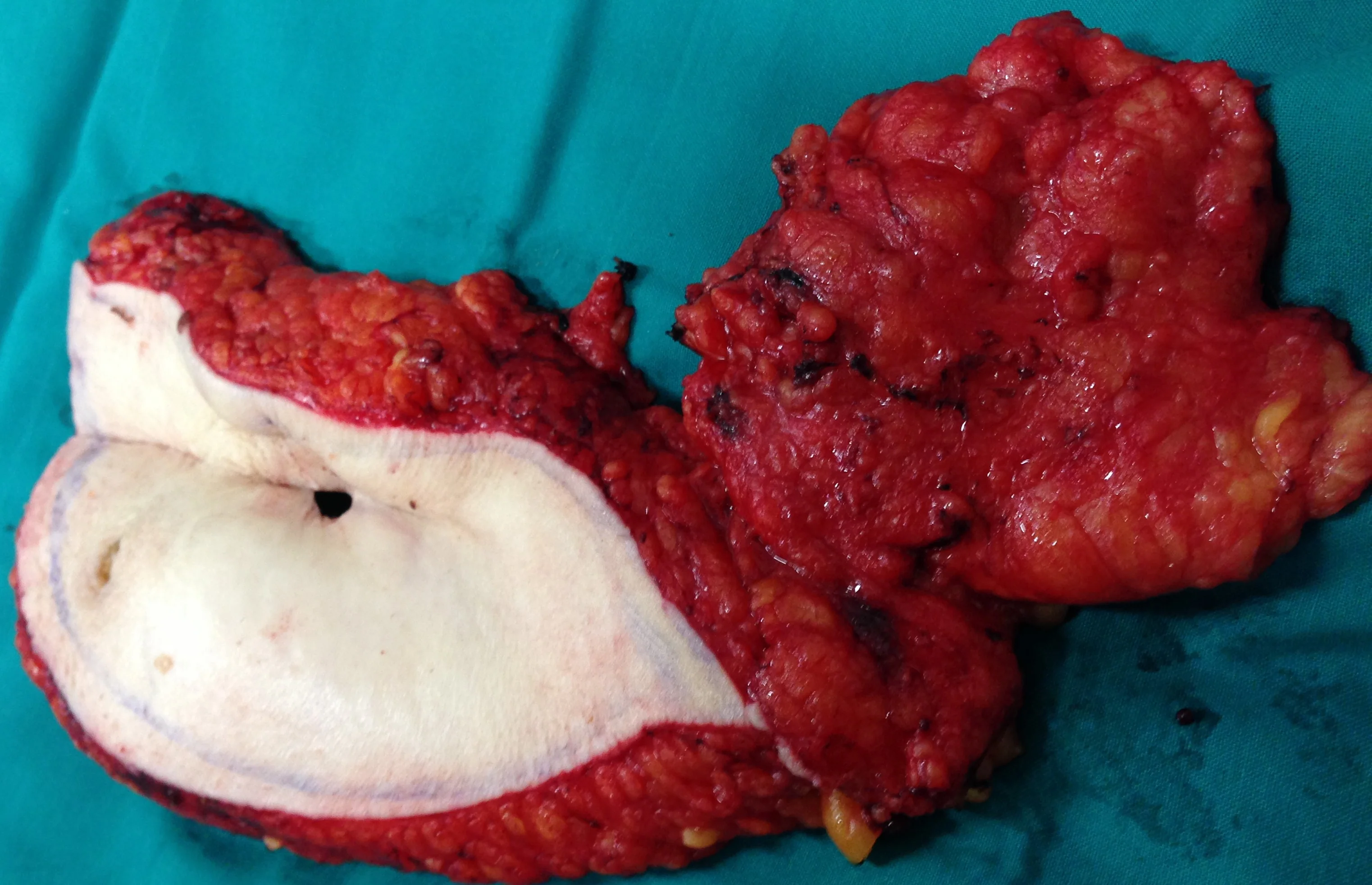
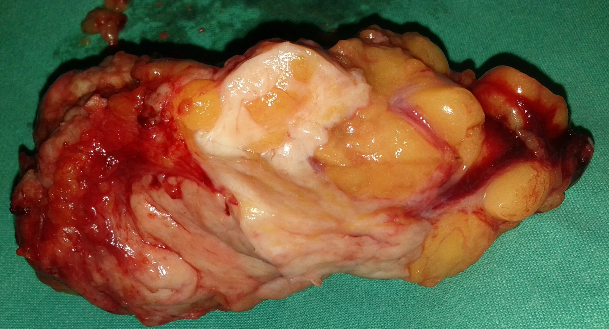
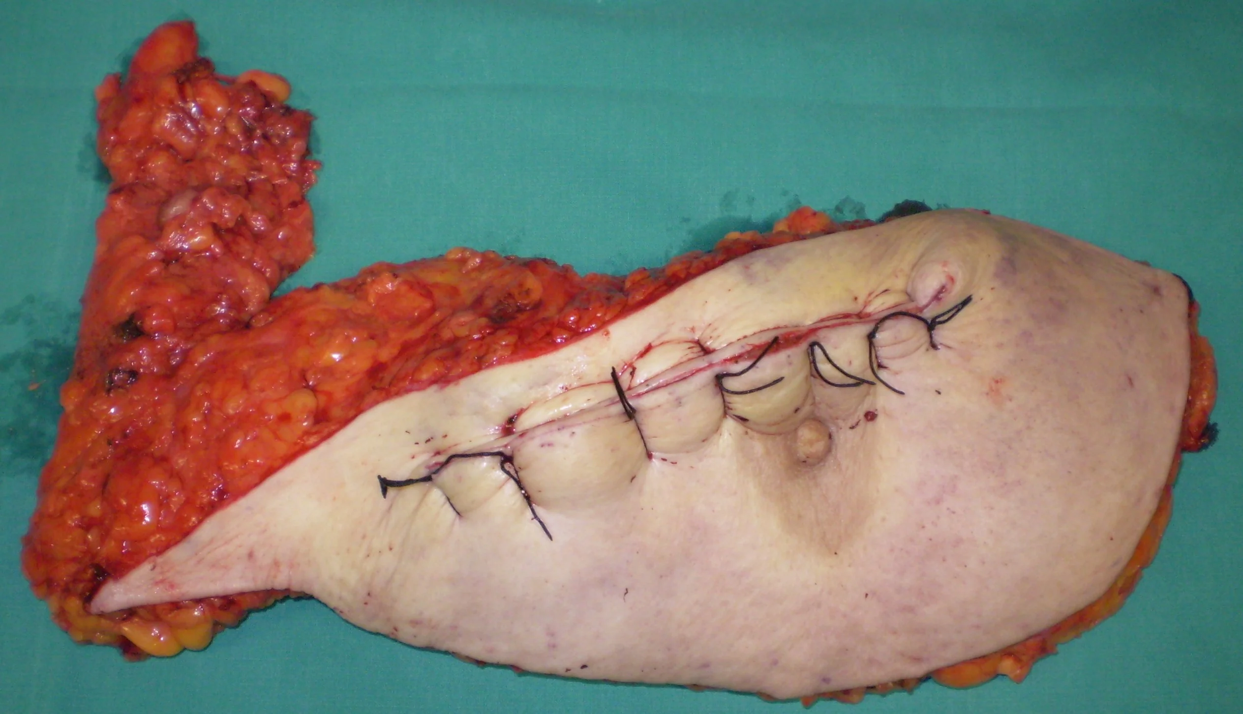
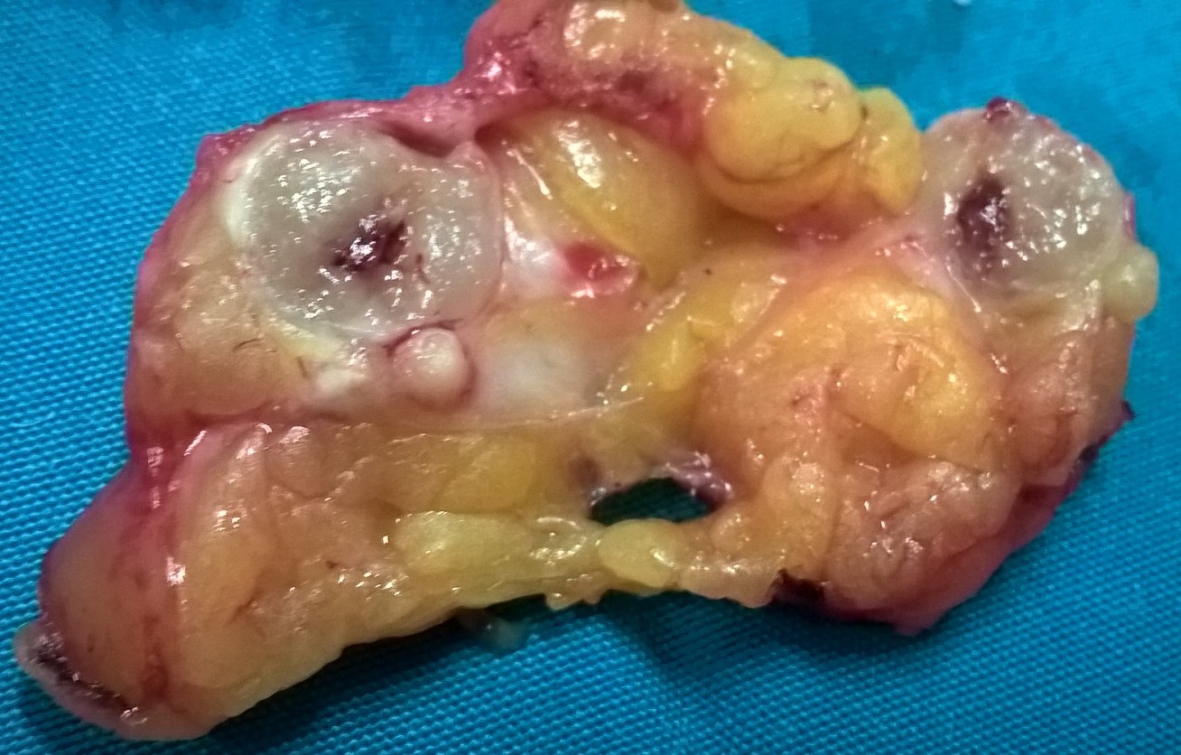
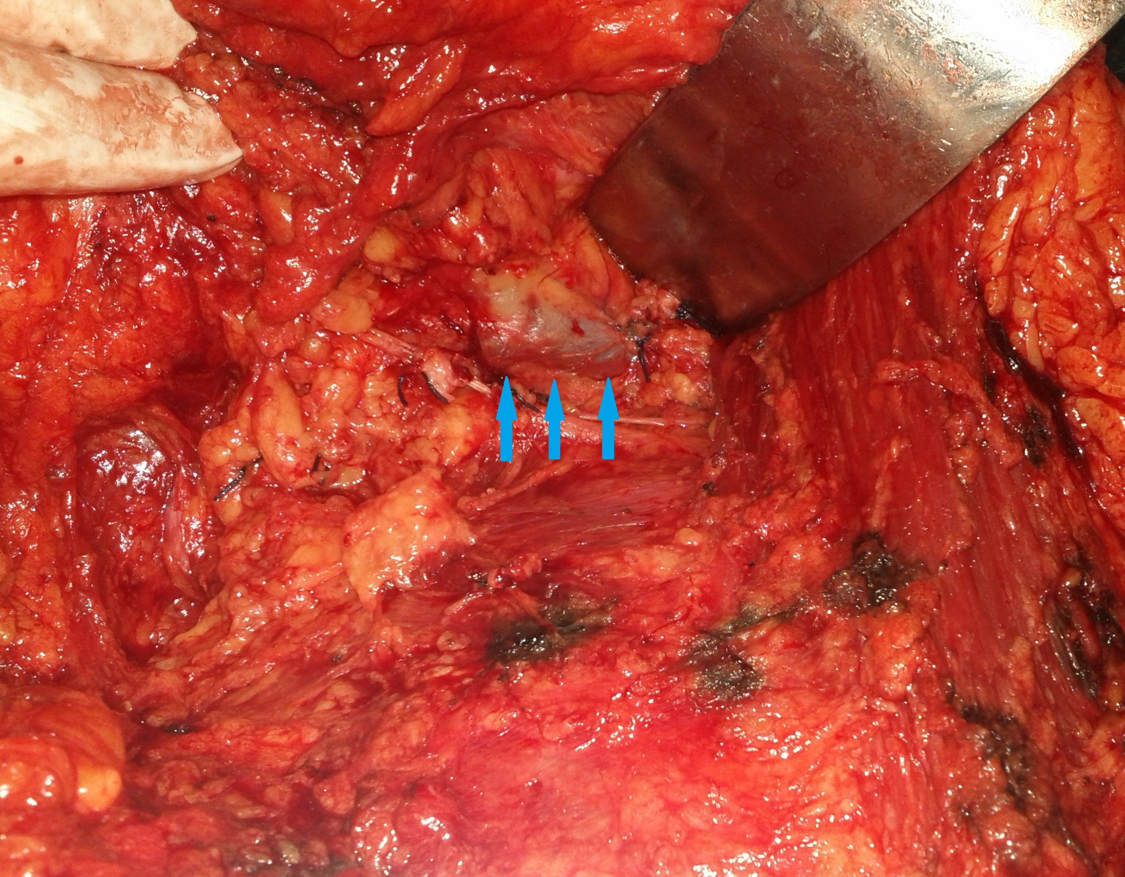
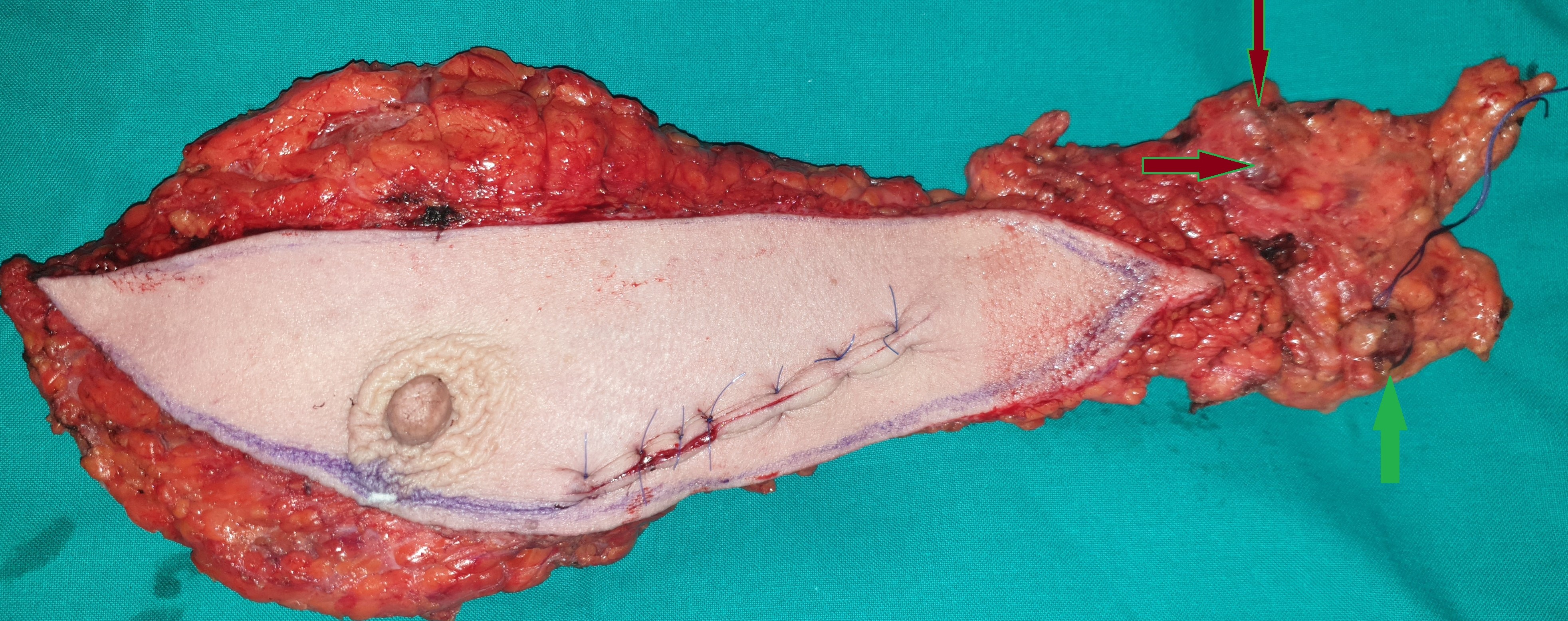
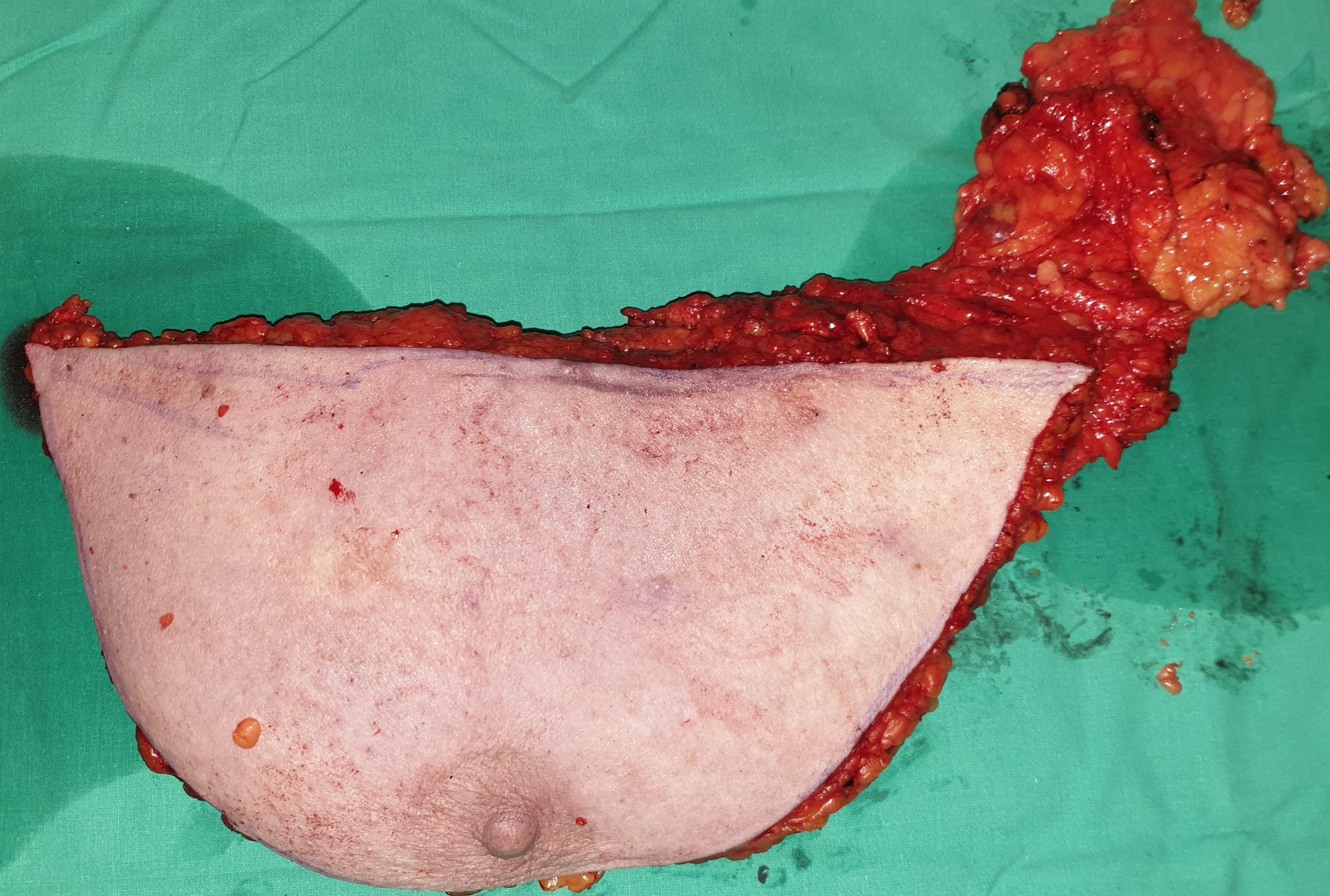
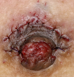
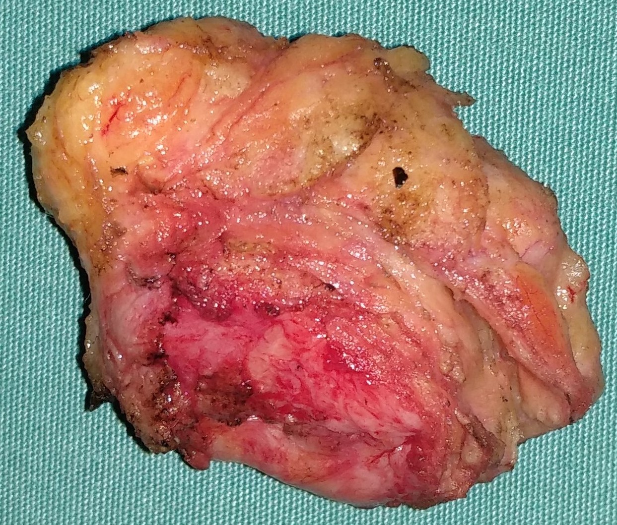
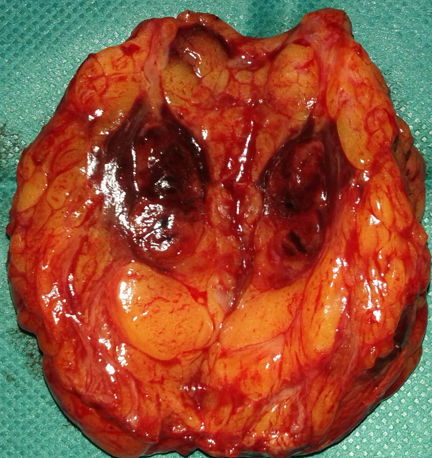
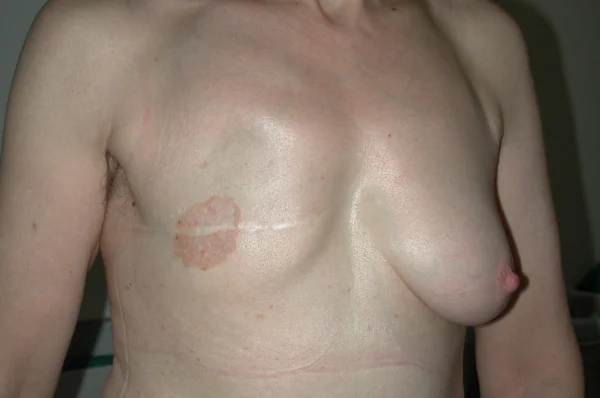
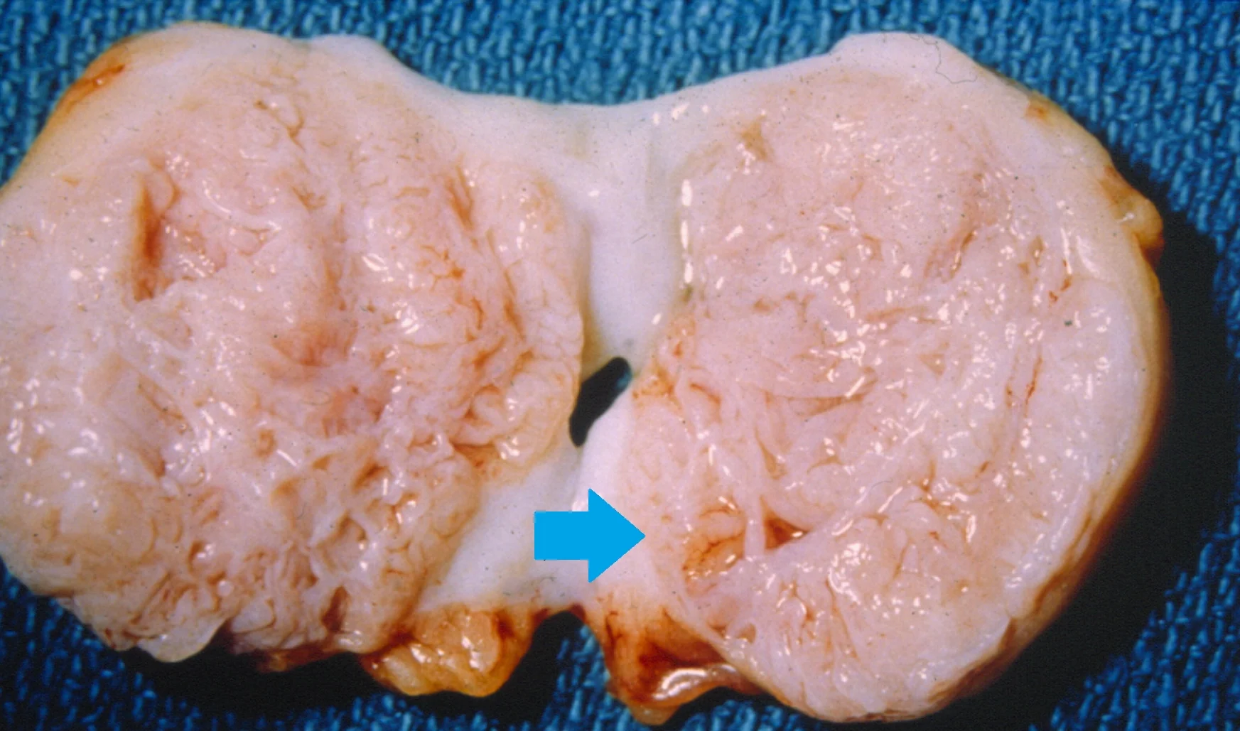
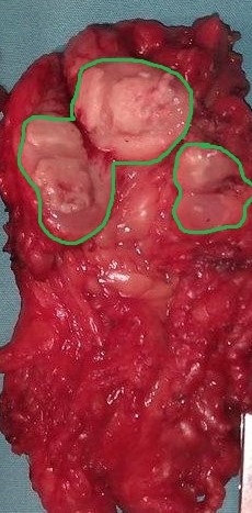
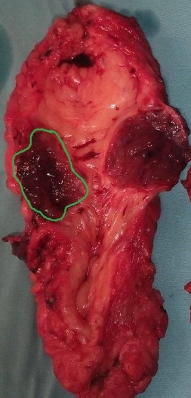
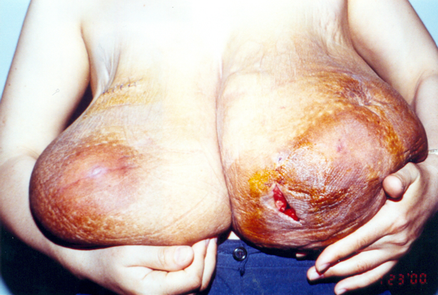
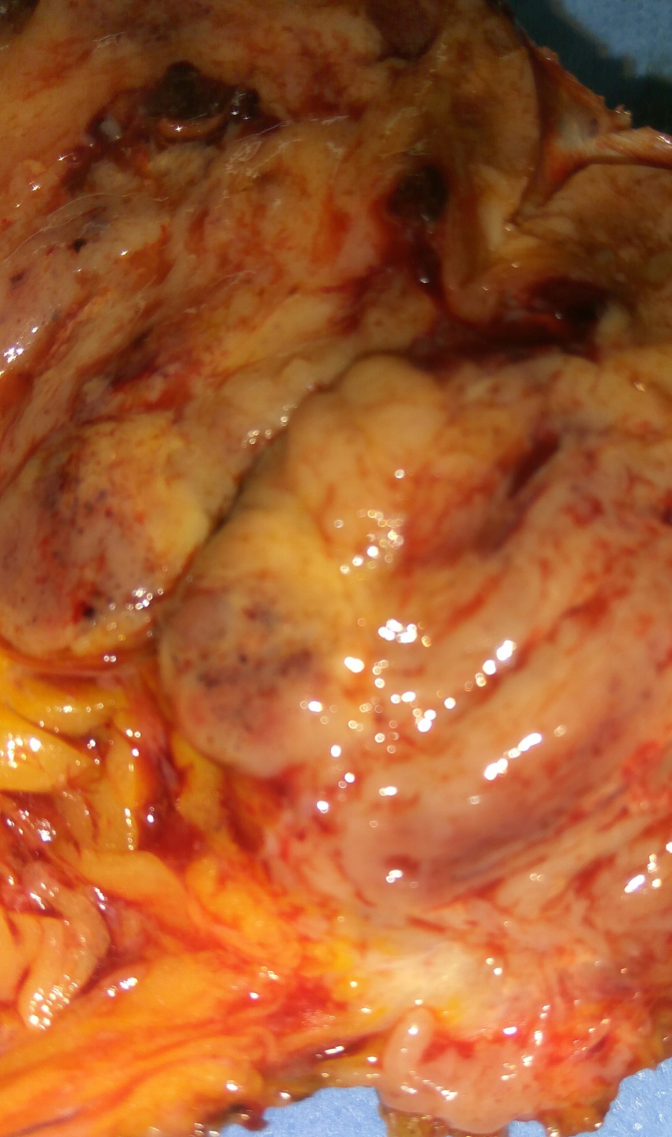
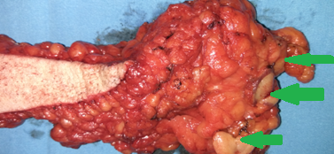
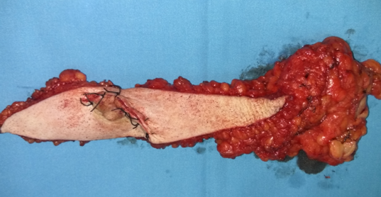
Surgical specimens of bilateral simple mastectomy and axillary lymph node dissection.(Courtesy Dr. V. Penopoulos).
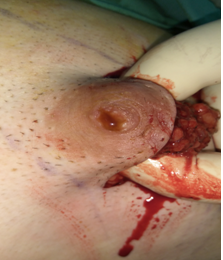
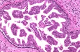
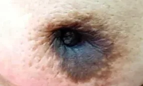
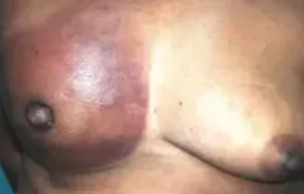
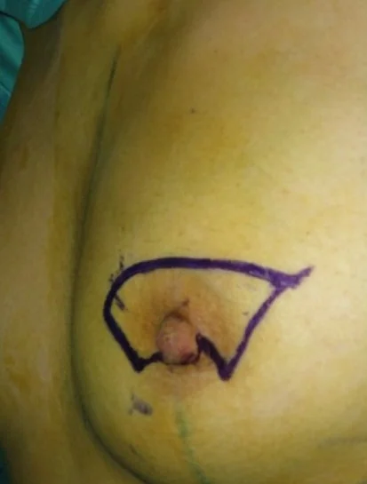
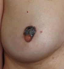
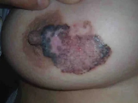
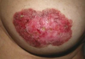
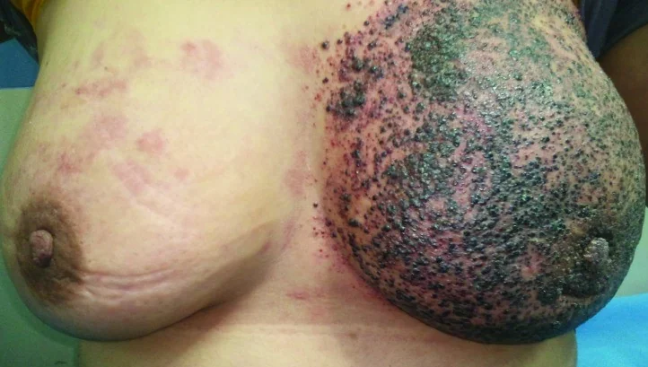
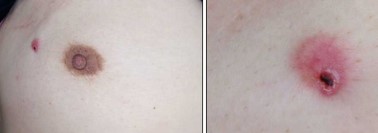
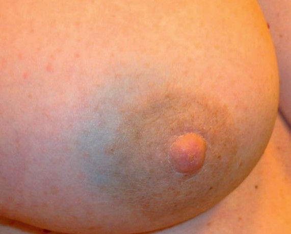
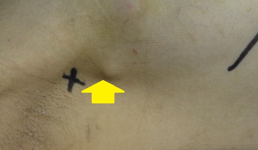
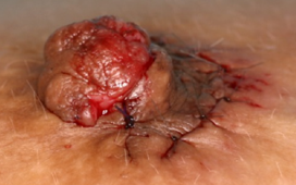
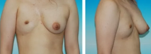
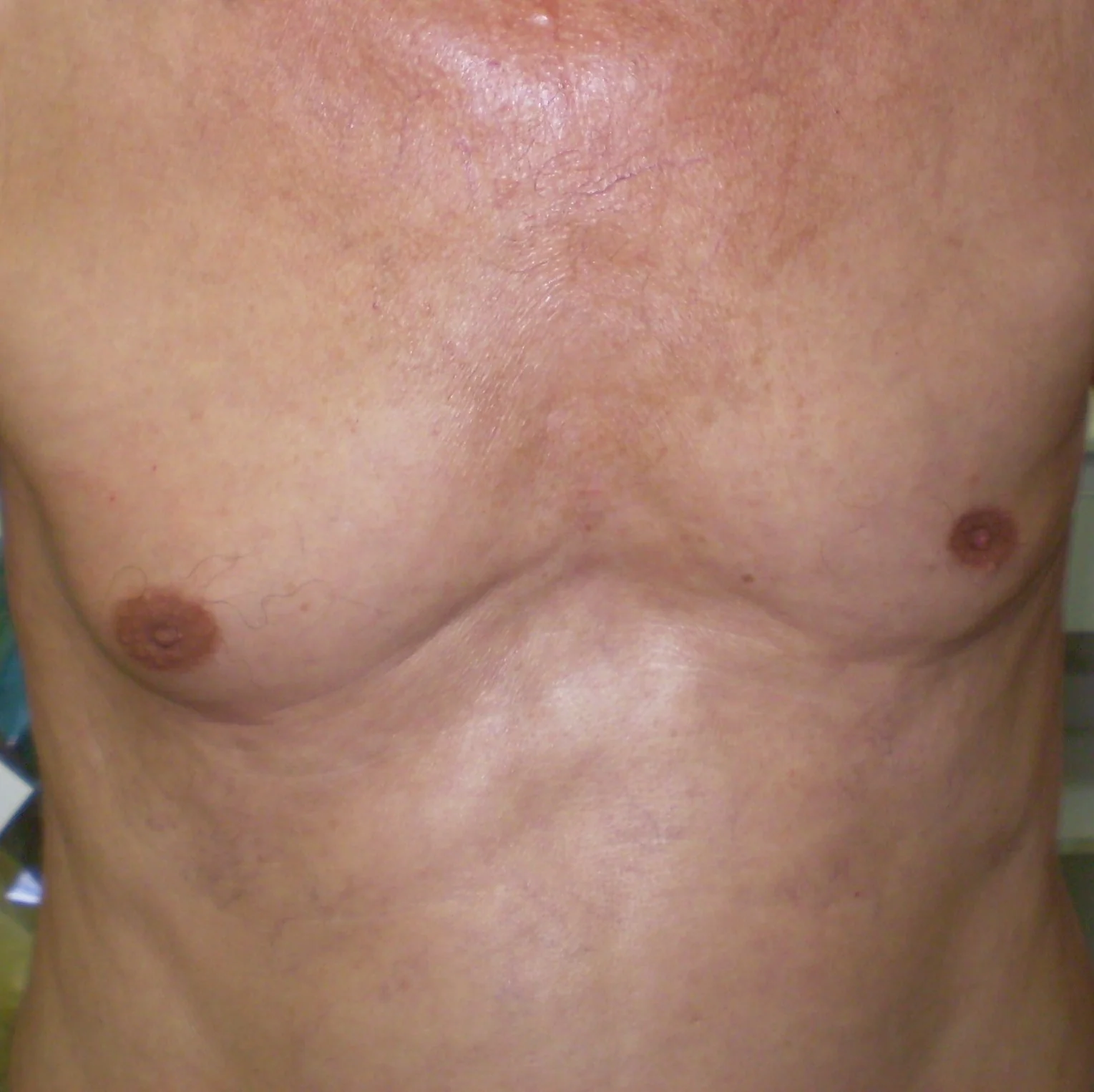
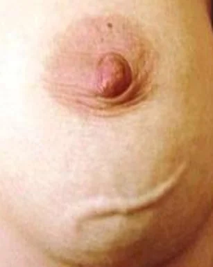
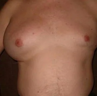
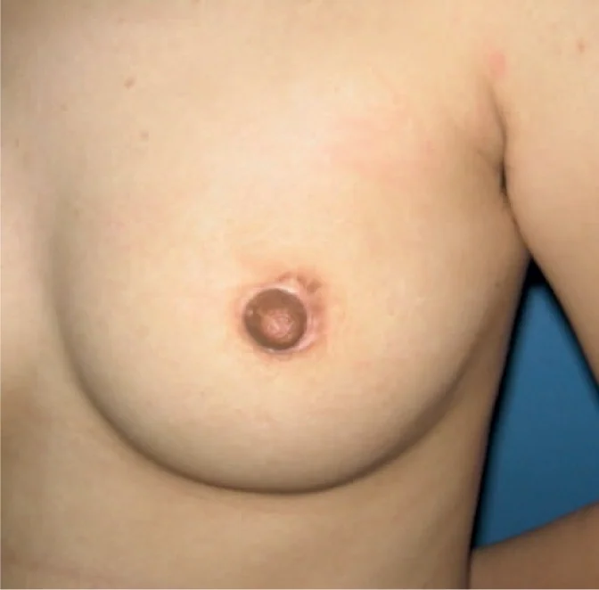
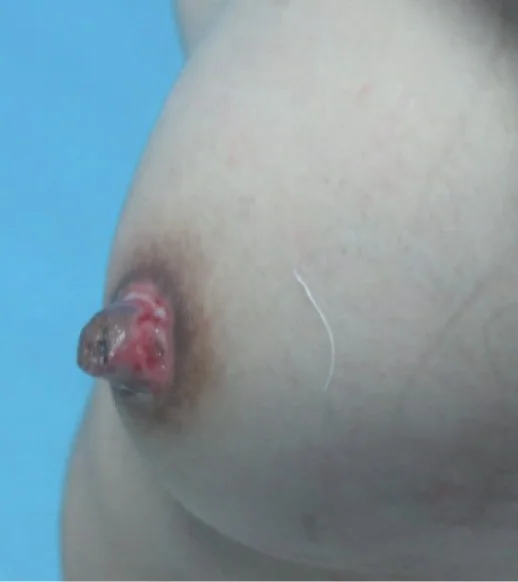
Bloody discharge from the nipple.Ductal carcinoma.(Courtesy Dr.V.Penopoulos).
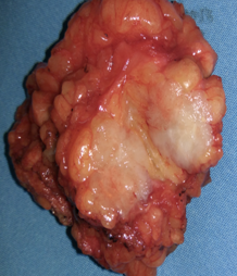
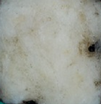
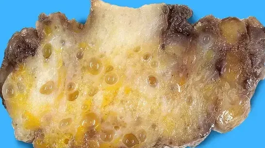
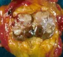
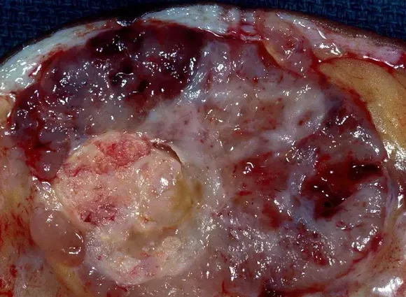
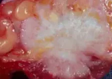
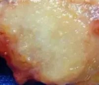
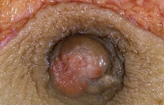
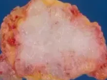
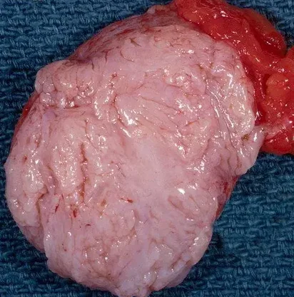
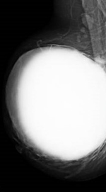
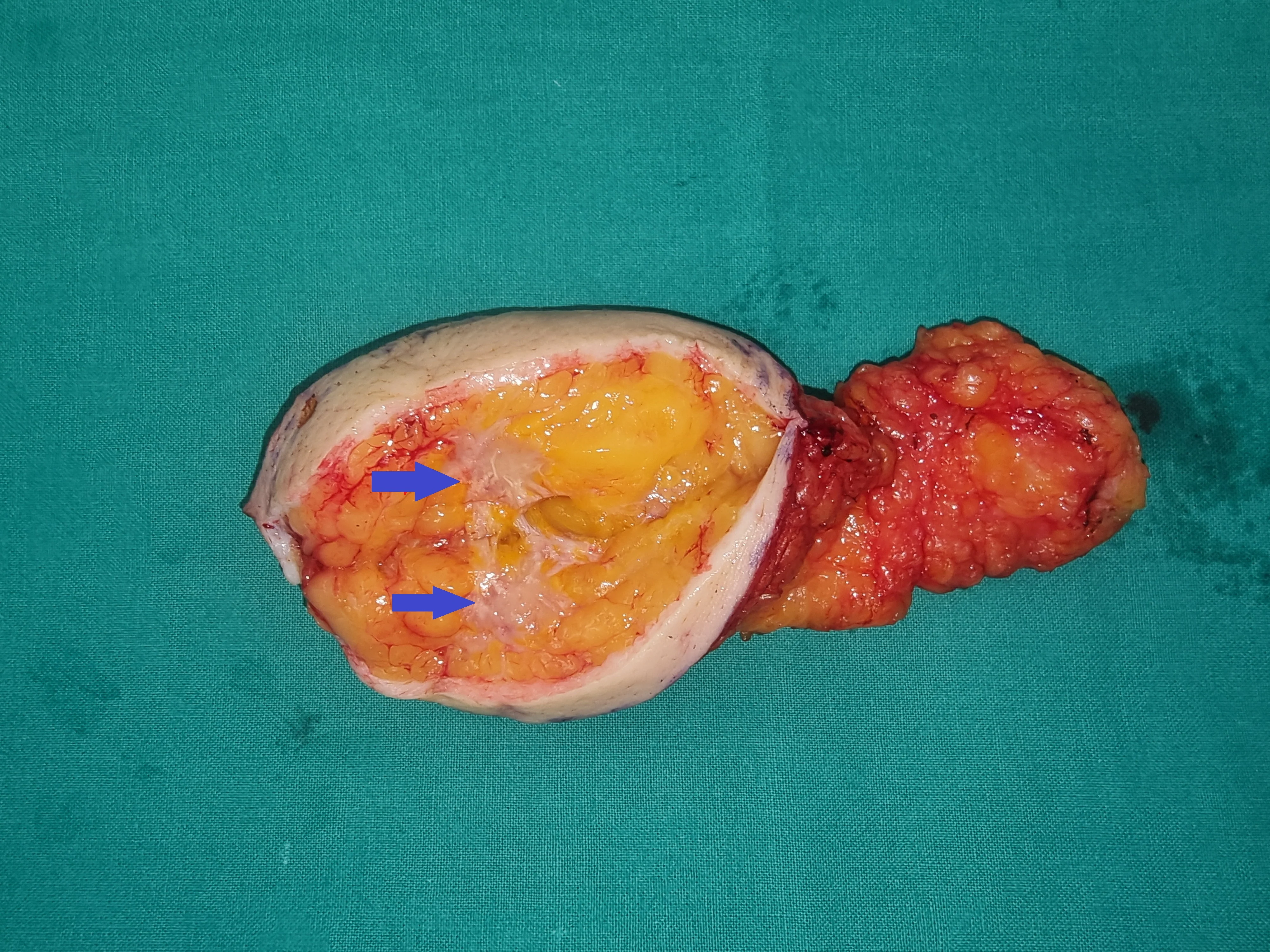
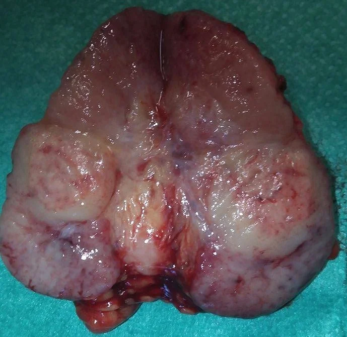
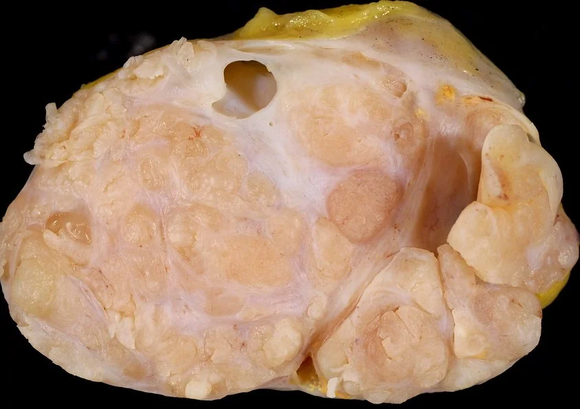
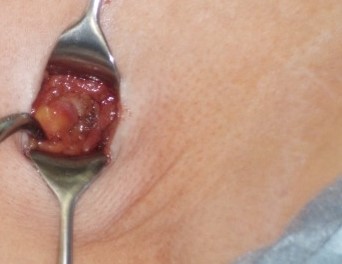
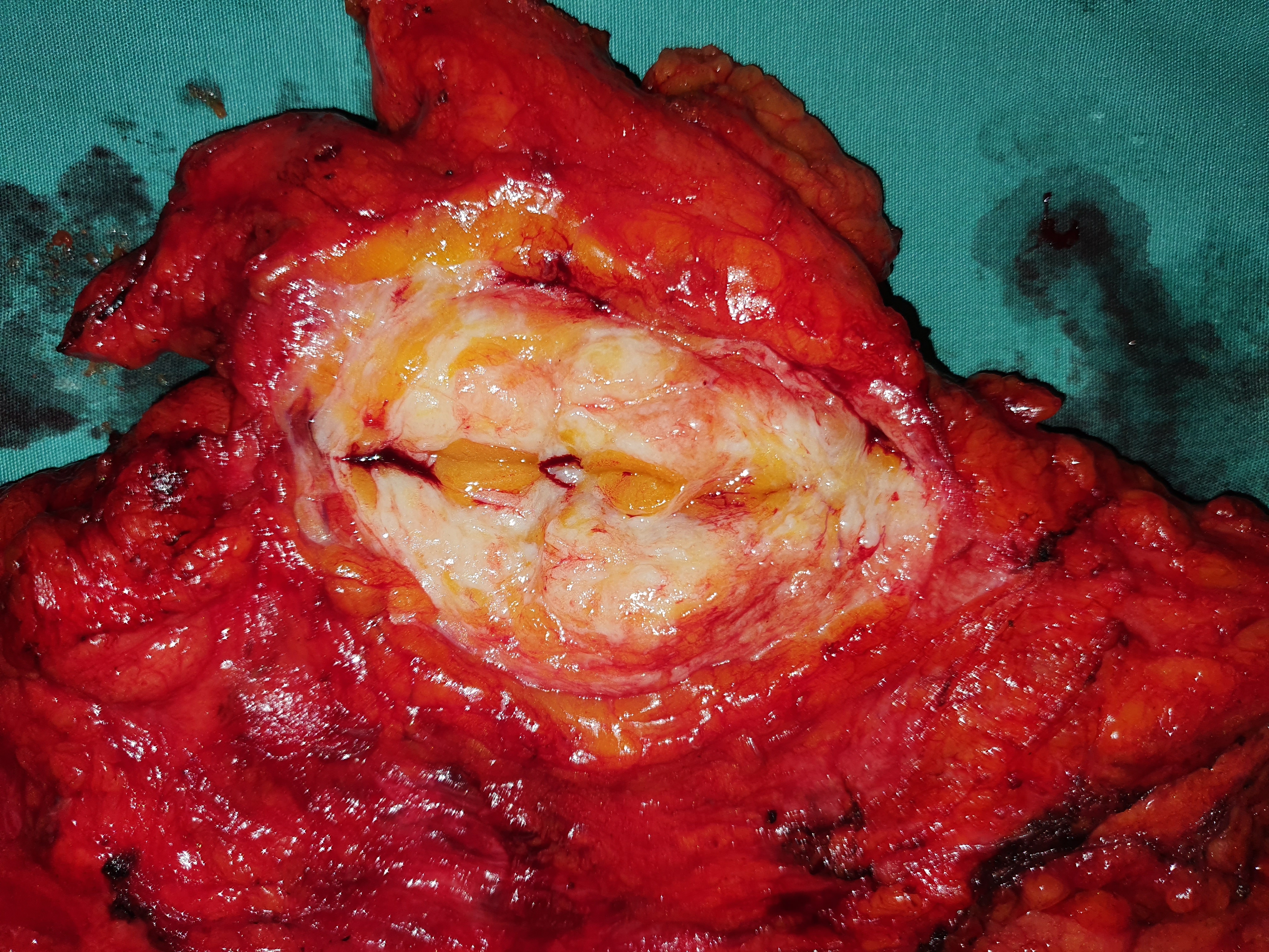
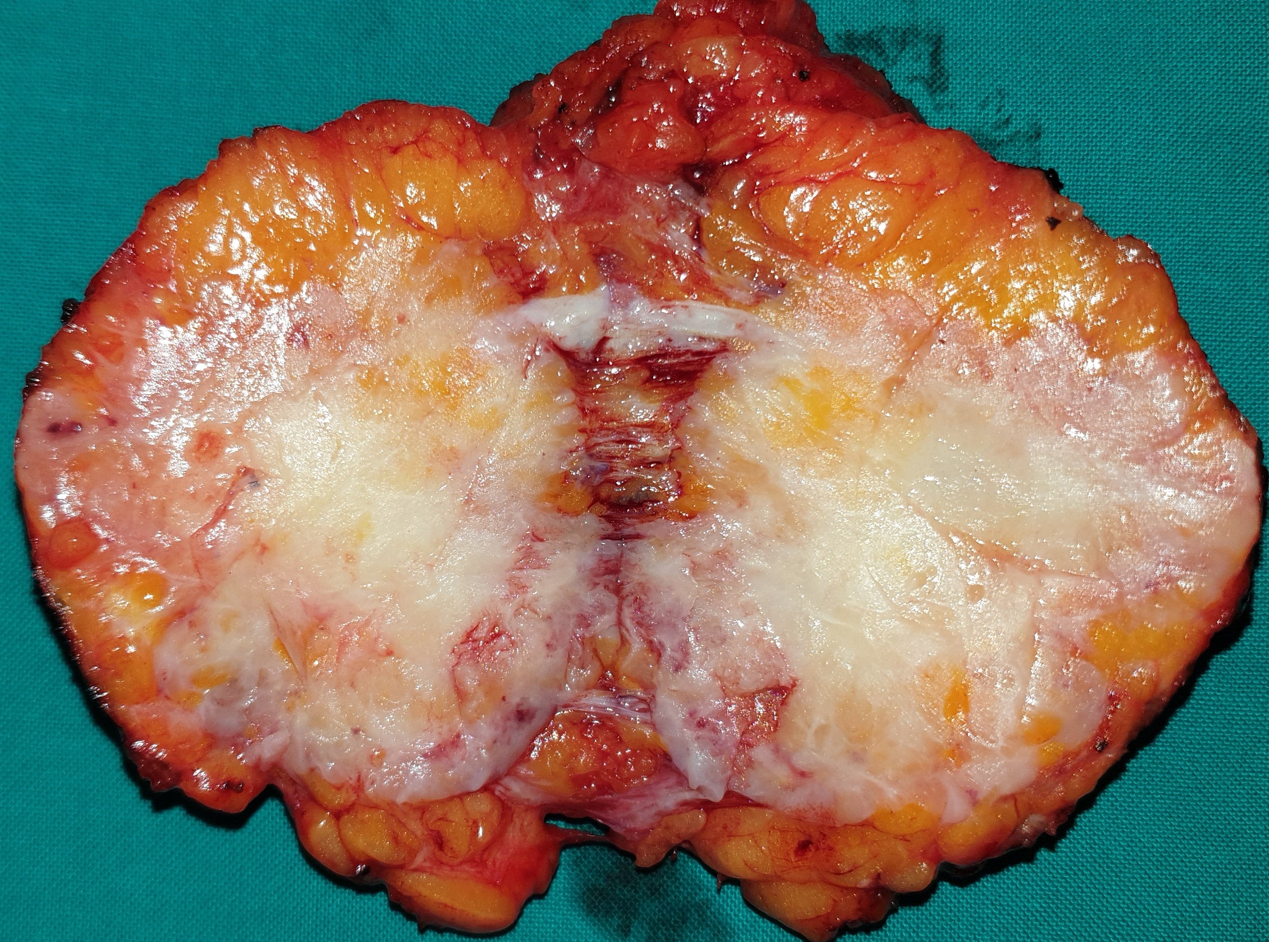
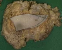
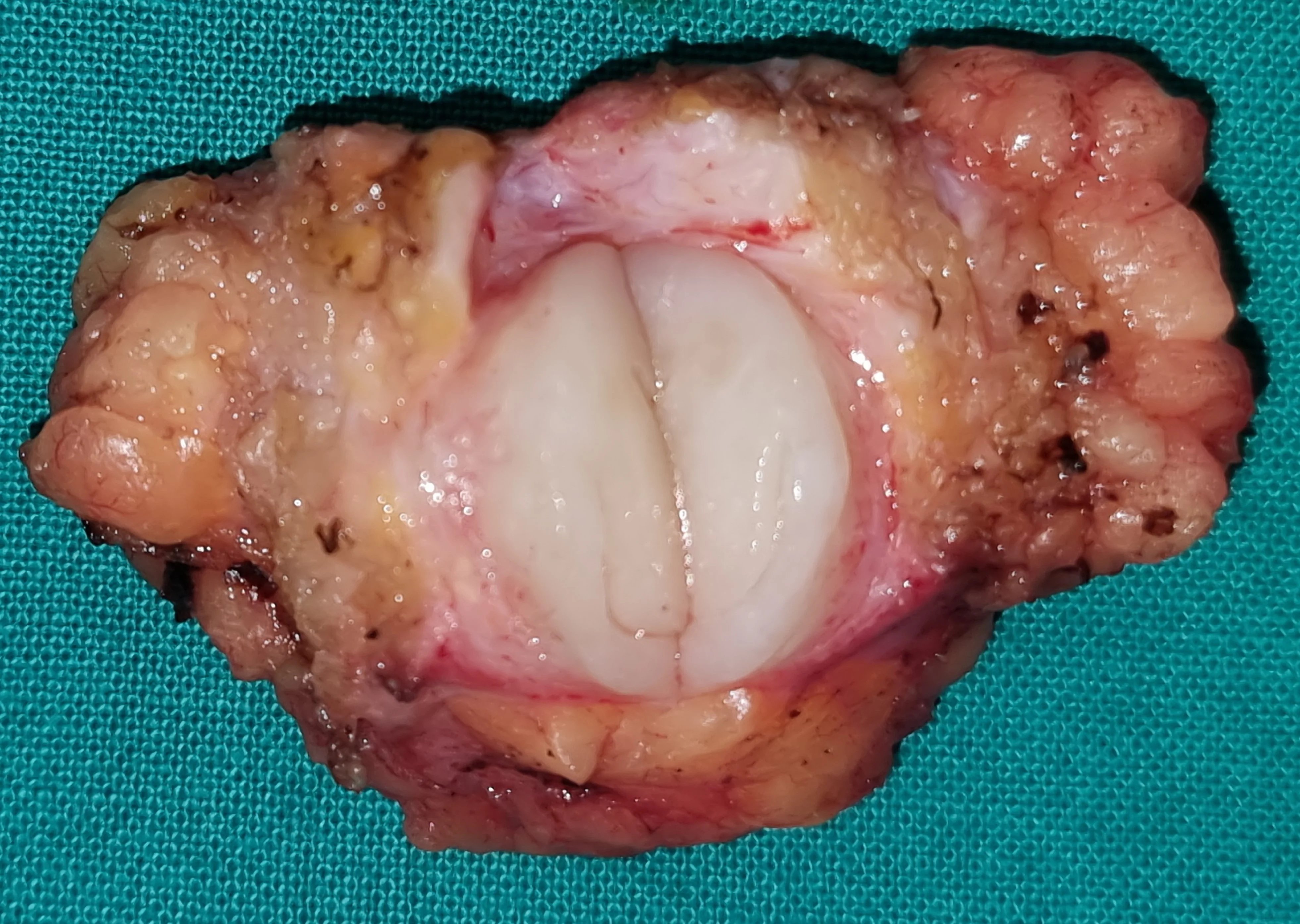
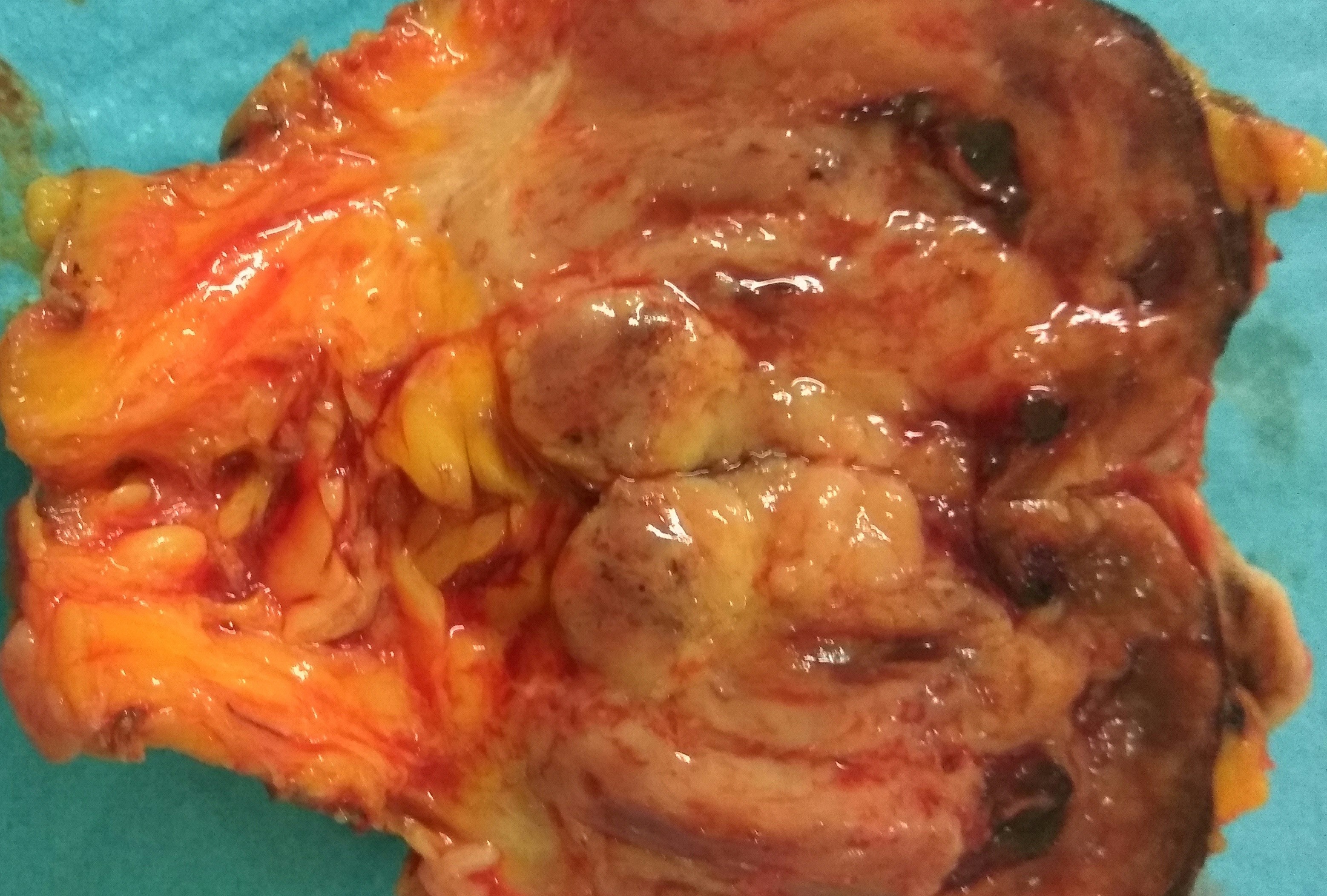
Excised nodule in cross-section. Its whitish color is apparent (Courtesy Dr. V. Penopoulos)
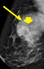
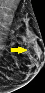
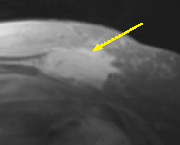
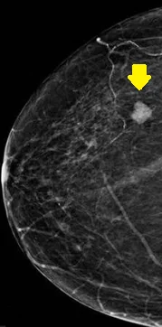
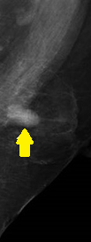
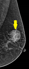
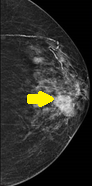
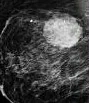
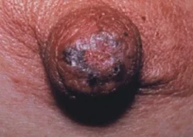
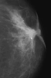
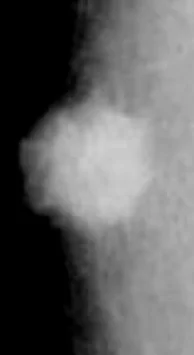
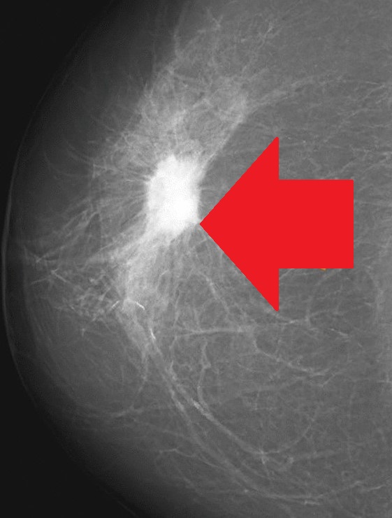
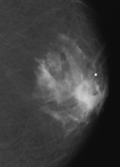
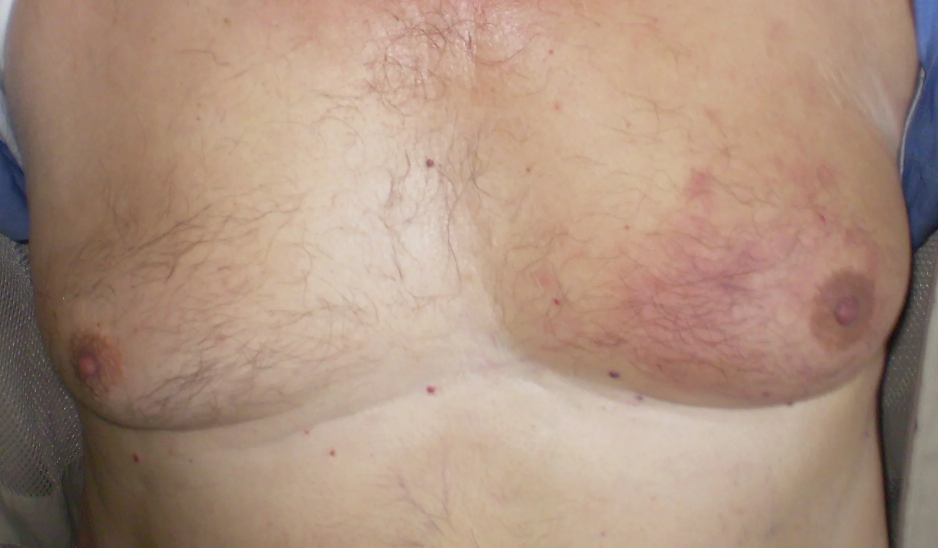
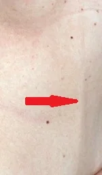
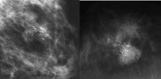
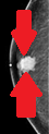
Digital mammography. Ductal carcinoma of the male breast (Courtesy Dr. V. Penopoulos)
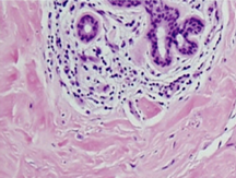
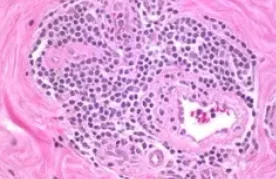
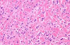
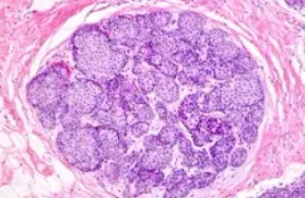
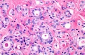
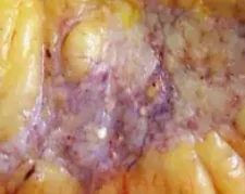
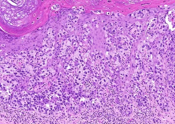
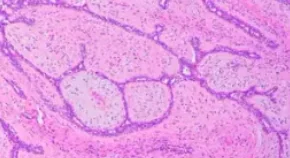
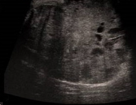
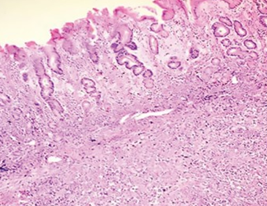
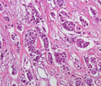
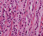
Marked stromal fibrosis and lymphocytic infiltration around the ducts of the breast gland (Courtesy Dr. V. Penopoulos)
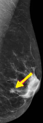
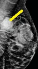
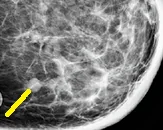
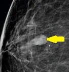
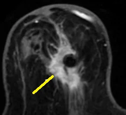
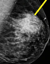
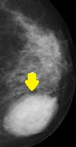
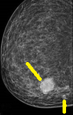
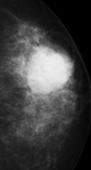
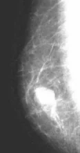
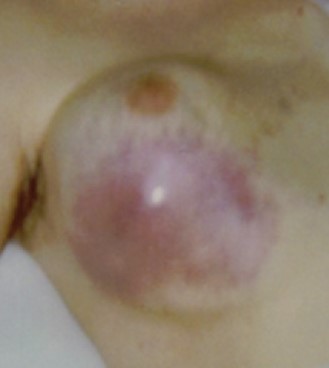
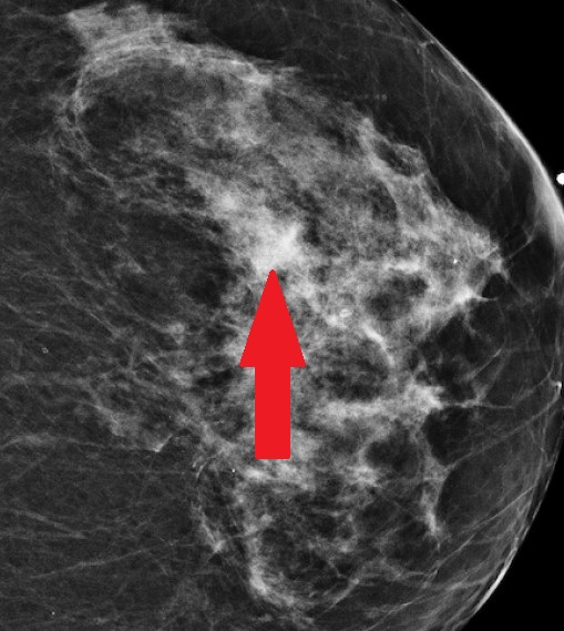
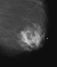
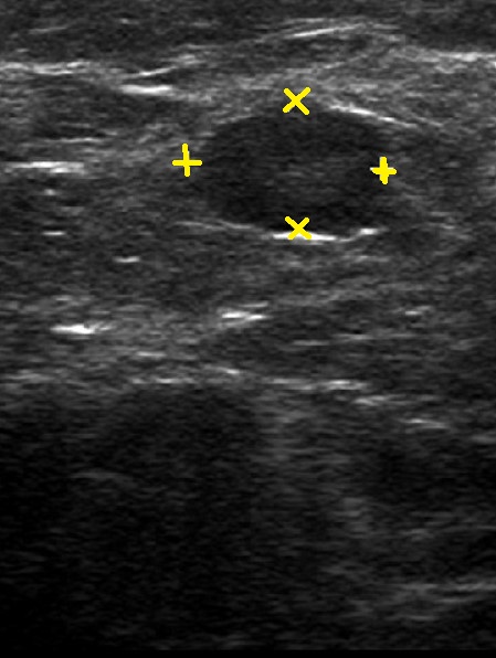
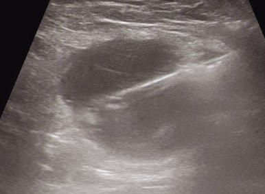
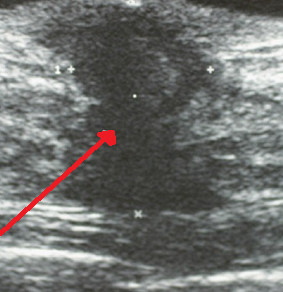
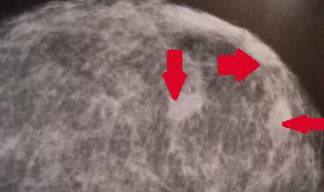
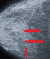
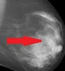
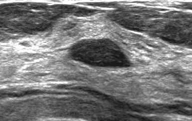
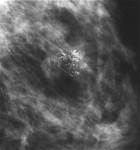
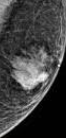
Left breast mammography.Heterogeneous dense fibroglandular pattern of the left breast parenchyma with the additional presence of a radiopaque nodule (Courtesy Dr. V. Penopoulos)
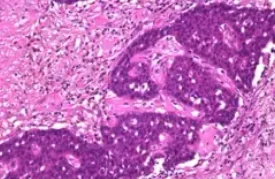
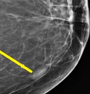
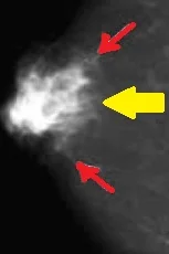
Digital mammography.Papillary carcinoma of the male breast.(Courtesy Dr.V.Penopoulos).