Περιτόναιο
30 images
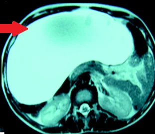
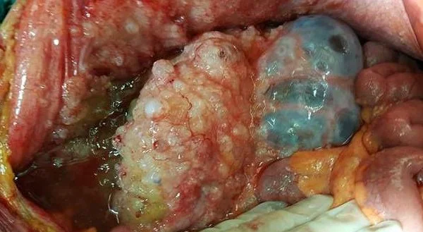
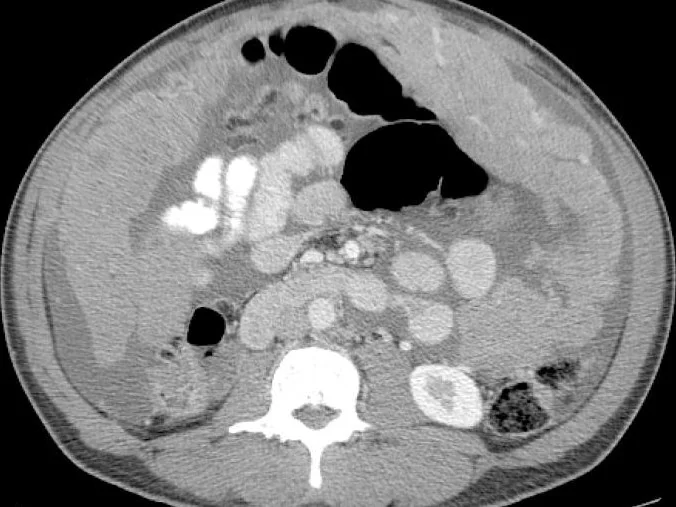
CYSTIC LYMPHANGIOMA
MALIGNANT MESOTHELIOMA
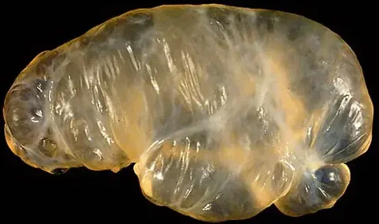
GIANT MESOTHELIAL CYST
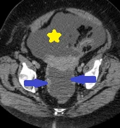
CYSTIC PERITONEAL LYMPHANGIOMA
CHEMICAL PERITONITIS
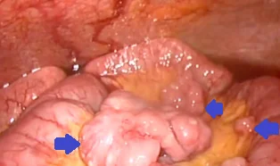
SOLITARY PERITONEAL CYST
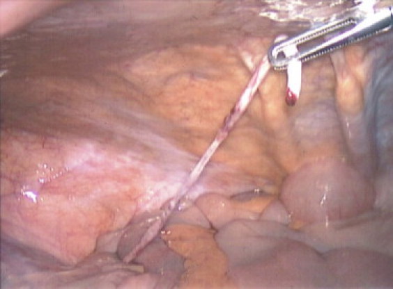
CONGENITAL BAND VITELLINE ARTERY
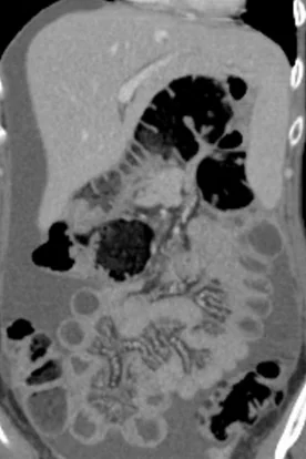
MALIGNANT PERITONEAL MESOTHELIOMA
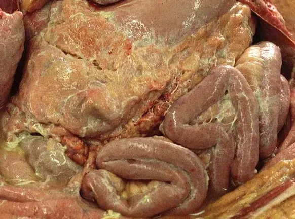
PERITONITIS
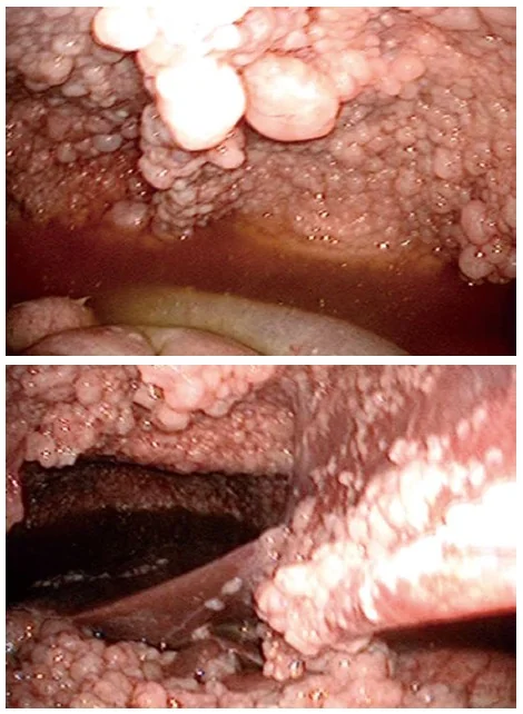
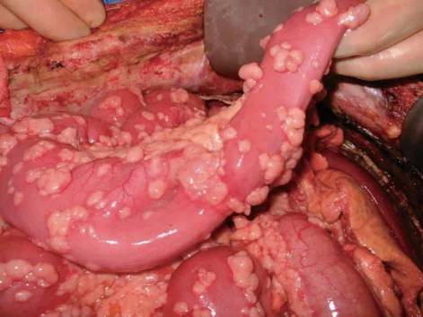
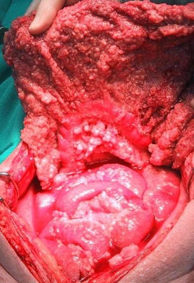
ILEAL NET - PERITONEAL DISSEMINATION
KRUKENBERG TUMOR
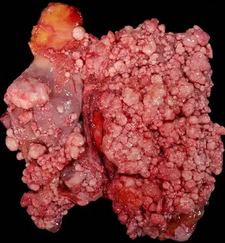
PSEUDOMYXOMA PERITONEI-APPENDICEAL MUCINOUS NEOPLASM
WELL DIFFERENTIATED PAPILLARY MESOTHELIOMA
PSEUDOMYXOMA PERITONEI
PRIMARY PERITONEAL SEROUS CARCINOMA
BENIGN MULTICYSTIC PERITONEAL MESOTHELIOMA
MALIGNANT SARCOMATOID PERITONEAL MESOTHELIOMA
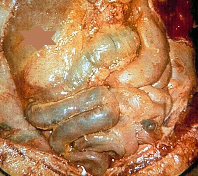
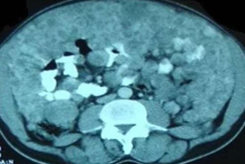
PERITONEAL CARCINOMATOSIS
PERITONEAL STROMAL TUMOR
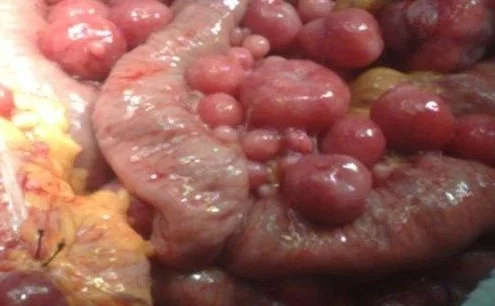
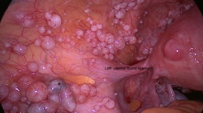
DISSEMINATED PERITONEAL LEIOMYOMATOSIS
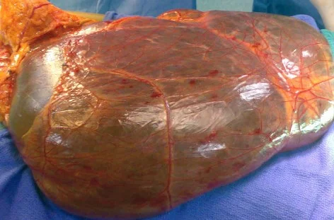
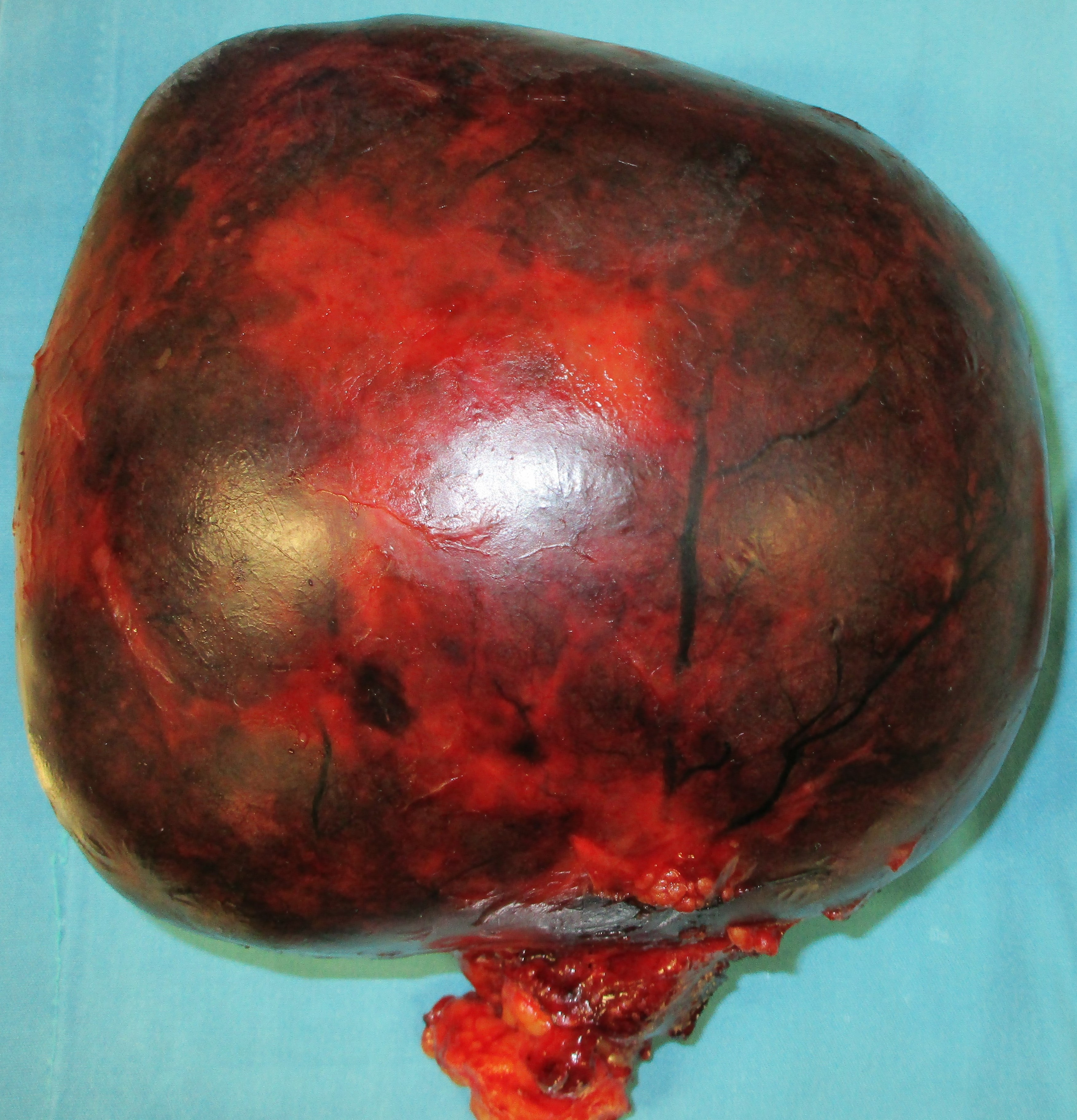
PRIMARY PERITONEAL TUMOR - SPECIMEN
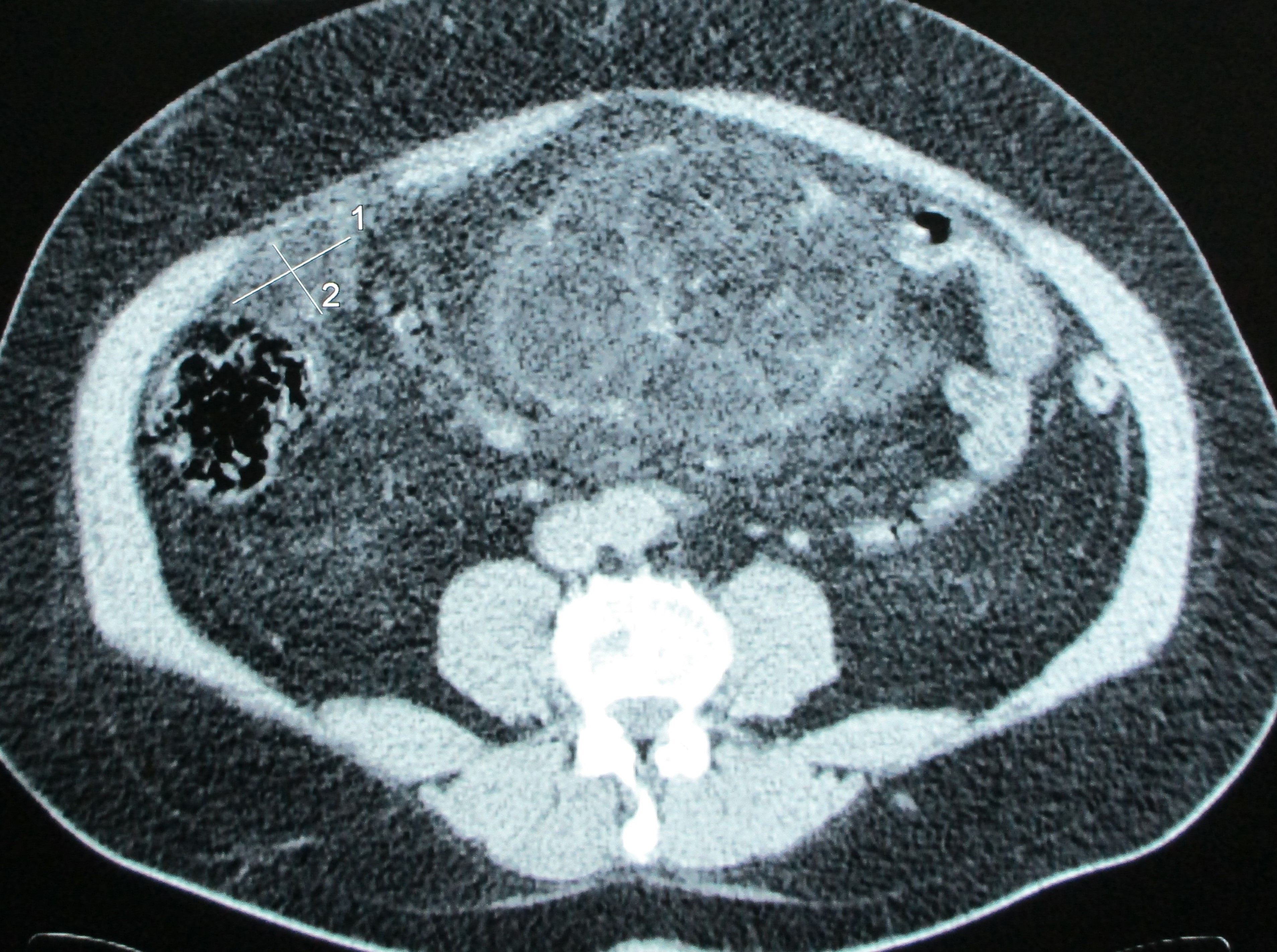
PRIMARY PERITONEAL TUMOR