Spleen
Σπλήνα
98 images
Upper abdominal CT scan 1 year after splenic autotransplantation. Enhancement of the transplanted splenic tissue after contrast injection is evident, demonstrating its viability and function (Courtesy Dr. V. Penopoulos)
Splenic graft within the omental pouch, secured to the parietal peritoneum in the left subdiaphragmatic area (Courtesy Dr. V. Penopoulos)
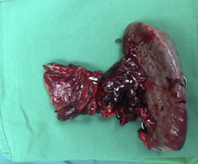
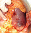
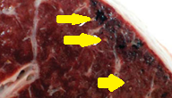
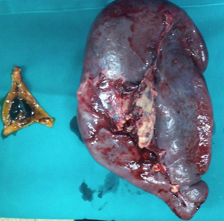
Distal pancreatectomy-splenectomy specimen, with the ruptured splenic artery aneurysm visible (Courtesy Dr. V. Penopoulos)
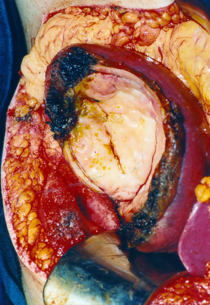
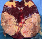
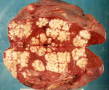
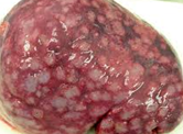
Epithelial splenic cyst. The trabeculated internal surface is visible, which may exhibit various types of epithelial lining (Courtesy Dr. V. Penopoulos)
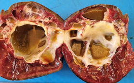
Epithelial splenic cyst. The trabeculated internal surface is visible, which may exhibit various types of epithelial lining (Courtesy Dr. V. Penopoulos)
Epithelial splenic cyst. The trabeculated internal surface is visible, which may exhibit various types of epithelial lining (Courtesy Dr. V. Penopoulos)
Epithelial splenic cyst. The trabeculated internal surface is visible, which may exhibit various types of epithelial lining (Courtesy Dr. V. Penopoulos)
Epithelial splenic cyst. The trabeculated internal surface is visible, which may exhibit various types of epithelial lining (Courtesy Dr. V. Penopoulos)
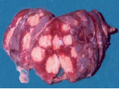
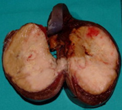
Surgical specimen of simple splenic cyst removal by partial splenectomy (Courtesy Dr. V. Penopoulos)
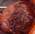
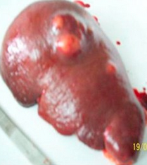
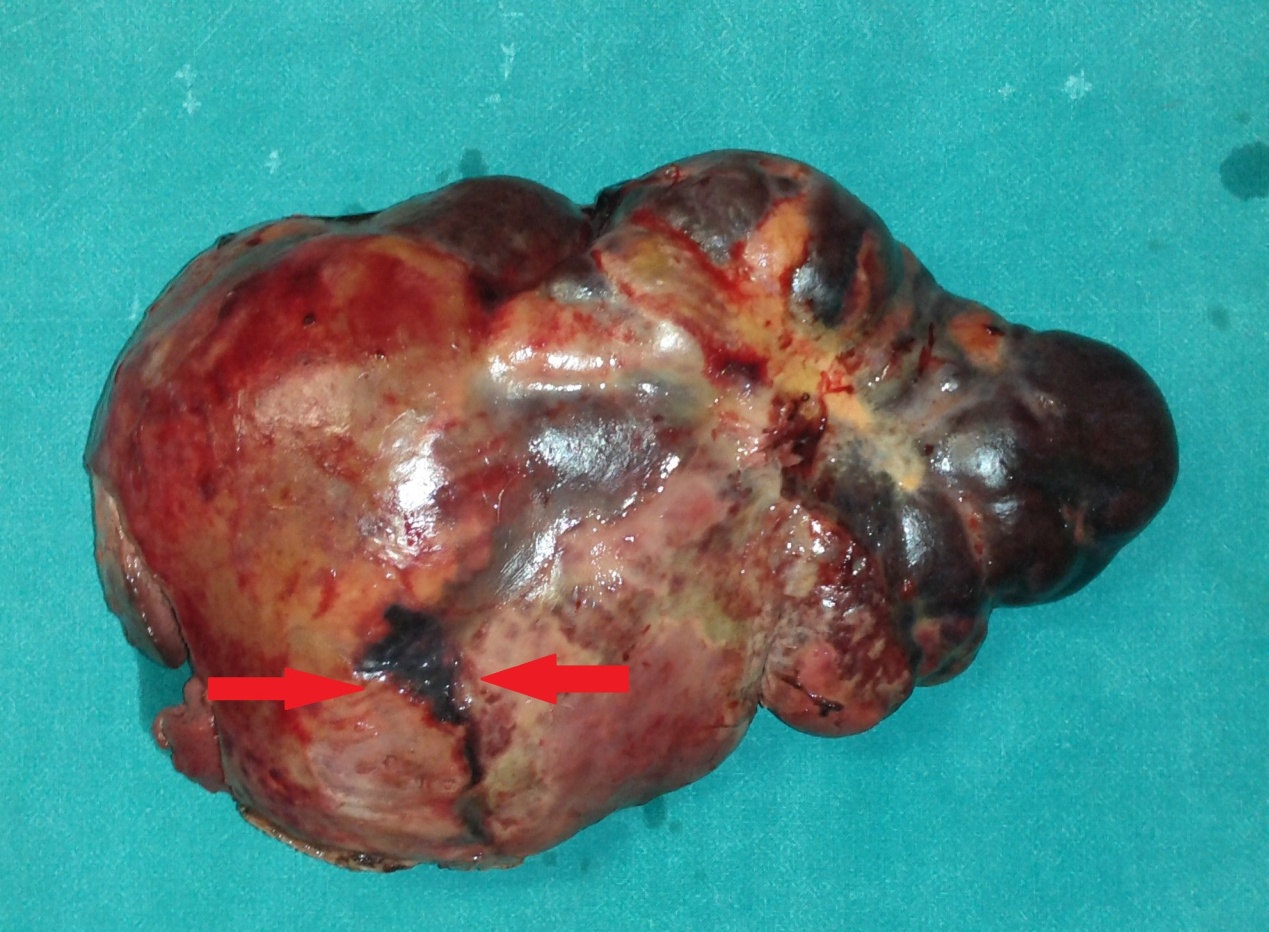
Macroscopic image of the splenectomy specimen. Littoral cell angioma (Courtesy Dr. V. Penopoulos)
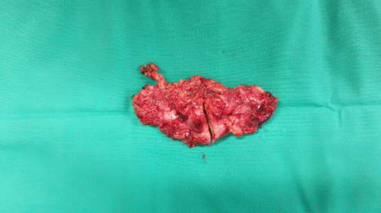
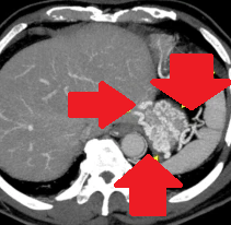
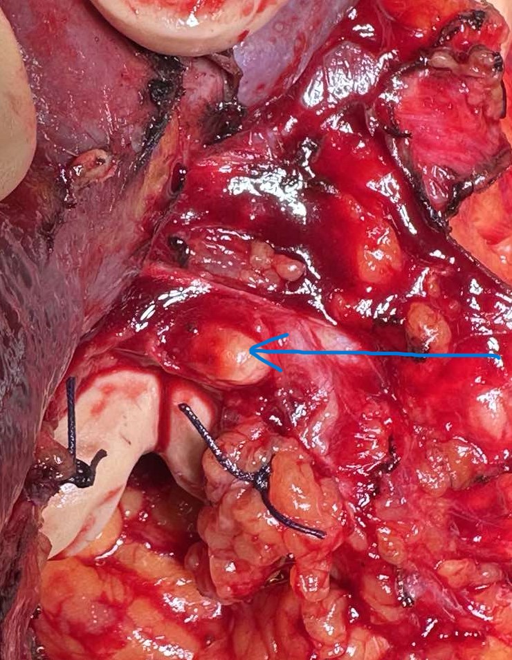
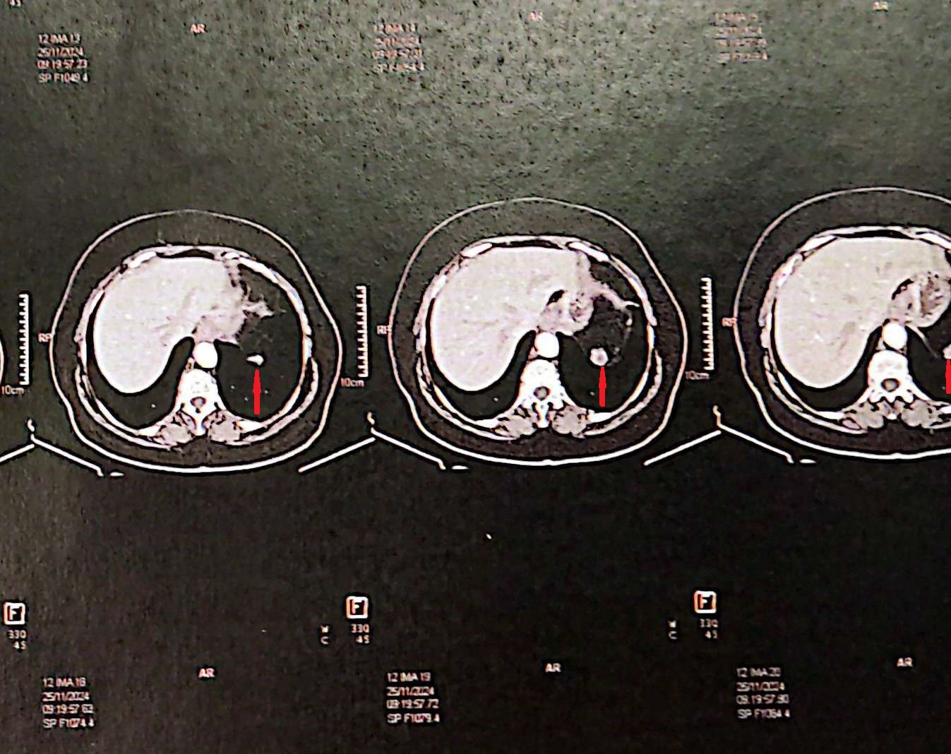
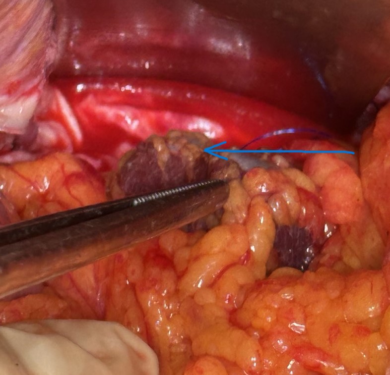
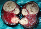
Excision of a metastatic implant for histopathological examination (Courtesy Dr. V. Penopoulos)

Excision of a metastatic implant for histopathological examination (Courtesy Dr. V. Penopoulos)
Excision of a metastatic implant for histopathological examination (Courtesy Dr. V. Penopoulos)
Excision of a metastatic implant for histopathological examination (Courtesy Dr. V. Penopoulos)
Excision of a metastatic implant for histopathological examination (Courtesy Dr. V. Penopoulos)
Excision of a metastatic implant for histopathological examination (Courtesy Dr. V. Penopoulos)
Excision of a metastatic implant for histopathological examination (Courtesy Dr. V. Penopoulos)
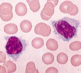
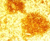
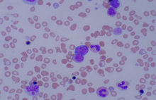
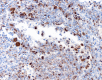
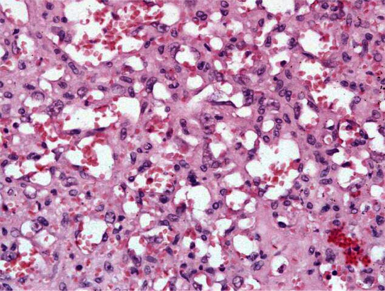
a) Splenic white pulp, enlarged with the presence of a large number of neoplastic cells (small and round). b) Villous lymphocytes, characteristic of SMZL (Courtesy Dr. V. Penopoulos)
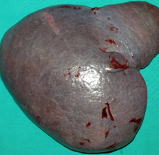
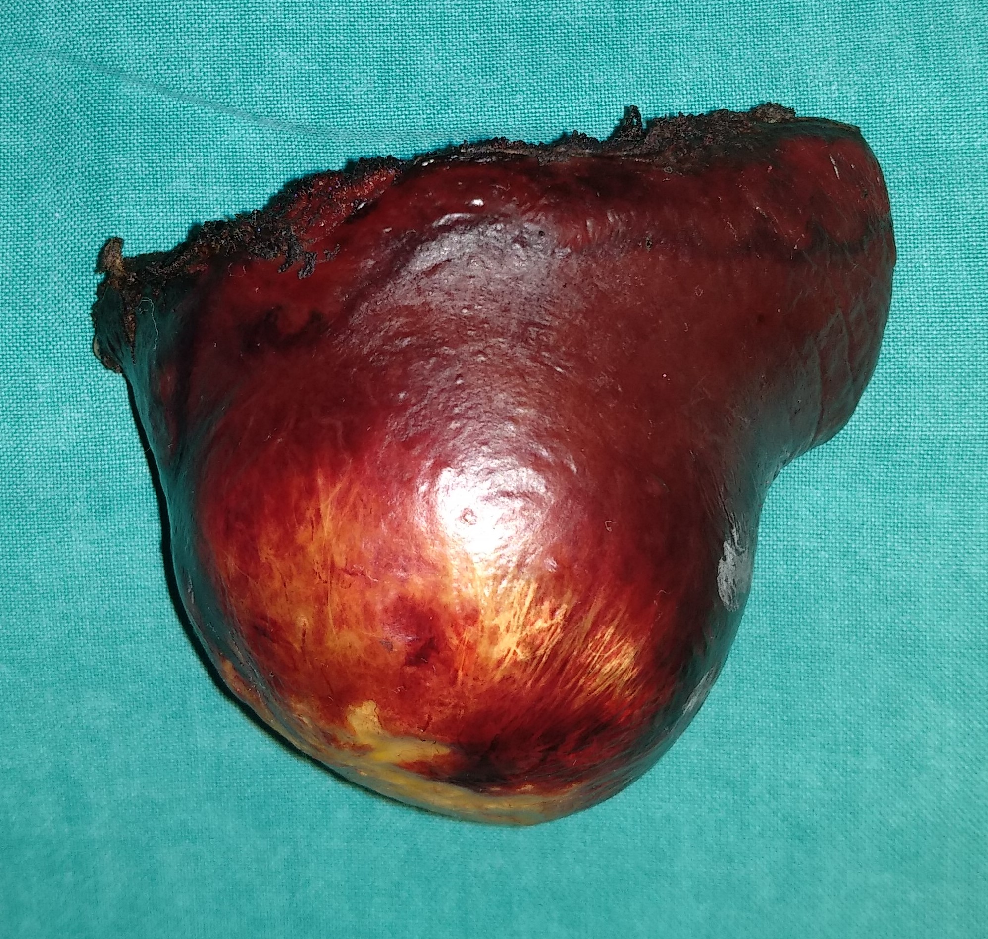
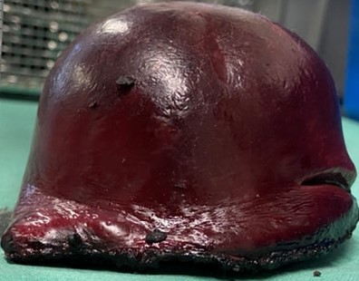
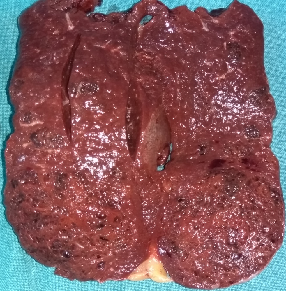
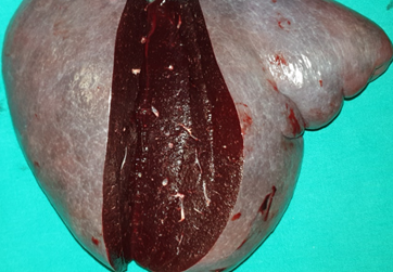
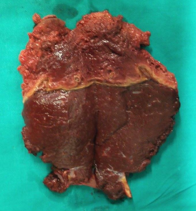
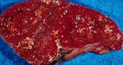
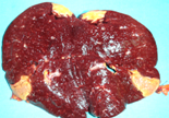
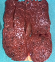
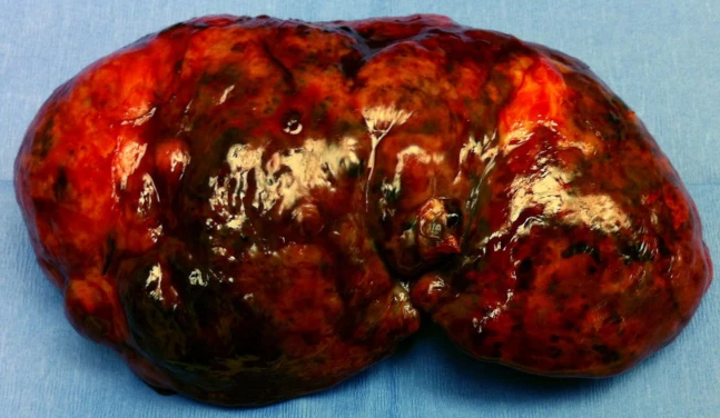
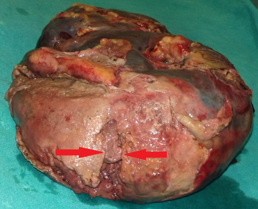
Splenectomy. Macroscopic image of the specimen. The dark red color of the splenic parenchyma is evident (Courtesy Dr. V. Penopoulos)


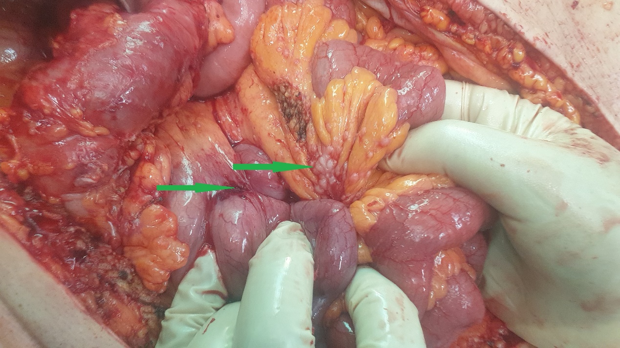
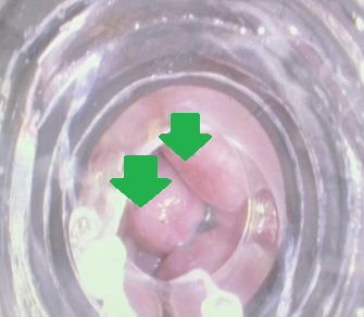
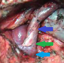
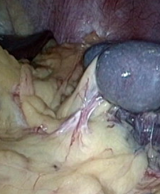
Green arrow – Splenic pedicle. Red arrow – Spleen fixed in retroperitoneal position. Courtesy Dr. V. Penopoulos.
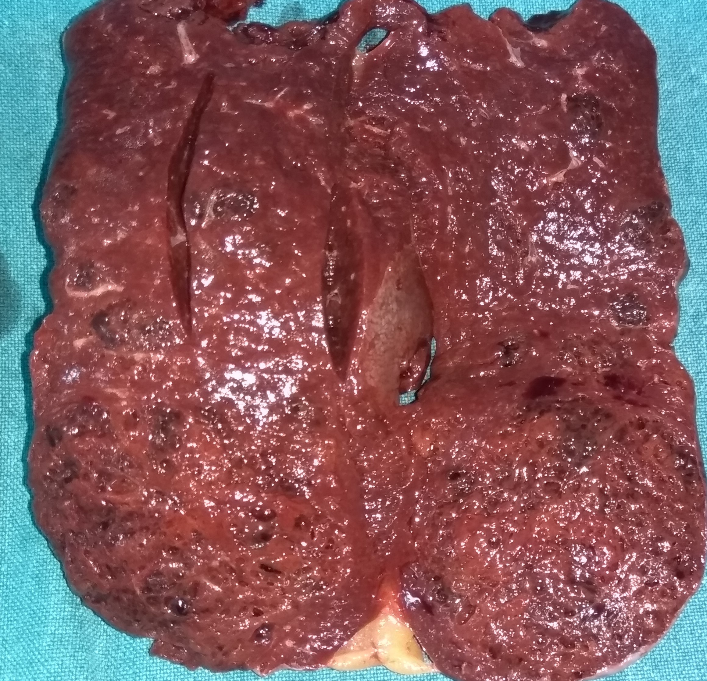
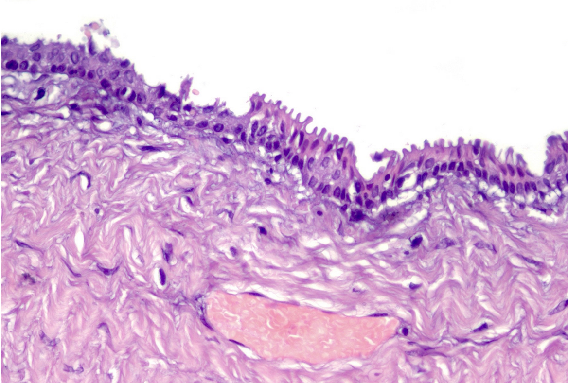
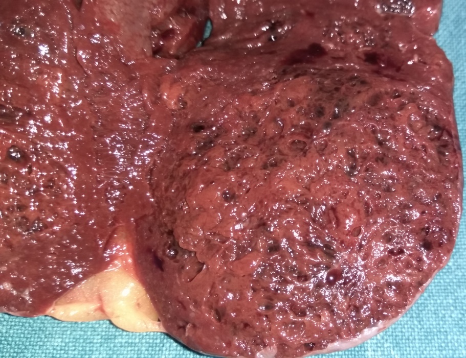
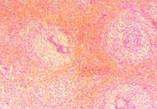
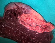
Figure 2 . The cut surface of the spleen , showing multiple sponge-like vascular spaces . ( Courtesy Dr . V . Penopoulos ) .
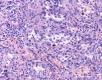
Figure 4 .Anastomosing channels lined by tall and plump cells, showing micropapillary architecture in some regions, but without nuclear atypia or mitotic activity. Deposition of hemosiderin can be seen in some cells. ( Courtesy Dr . V . Penopoulos ) .
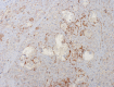
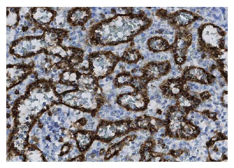
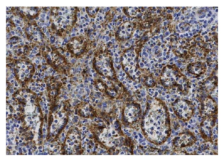
Figure 8 . The anastomosing channels lined cells express antigen FVIII. ( Courtesy Dr . V . Penopoulos ) .
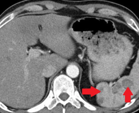
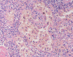
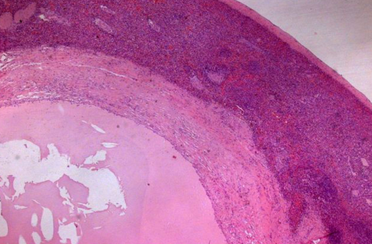
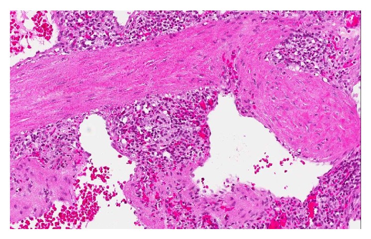
Figure 3 . Large sinus-like anastomosing channels, with deciduous endothelial cells in the channel. ( Courtesy Dr . V . Penopoulos ) .
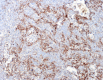
Figure 5 . The anastomosing channels lined cells express antigen CD31. (Courtesy Dr . V . Penopoulos ) .
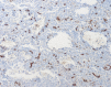
Figure 6 . Endothelium of peripheral vessels express antigen CD34, but not the anastomosing channels lined cells. ( Courtesy Dr . V . Penopoulos ) .
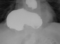
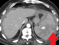
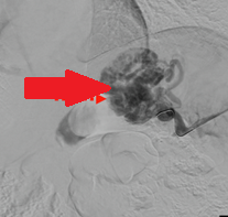
Lower sections of abdominal computed tomography – Red arrow: Wandering spleen torsion. Courtesy Dr. V. Penopoulos.
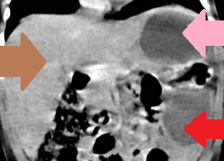
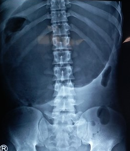
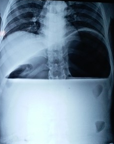
Red arrow – Wandering spleen. Pink arrow – Gastric volvulus. Brown arrow – Liver. Courtesy Dr. V. Penopoulos.
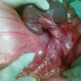
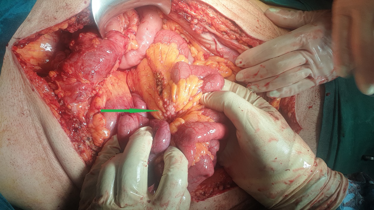
Green arrow – Splenic pedicle. Red arrow – Spleen fixed in retroperitoneal position. Courtesy Dr. V. Penopoulos.
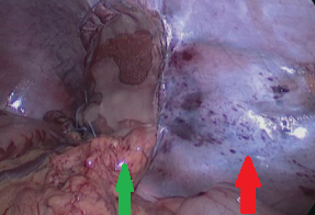
Green arrow – Splenic pedicle. Red arrow – Spleen fixed in retroperitoneal position. Courtesy Dr. V. Penopoulos.
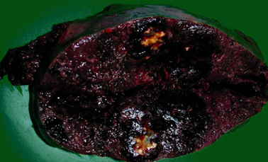
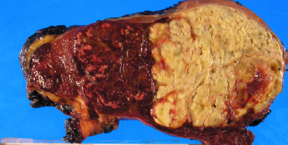
The enlarged spleen in cross-section – dark-colored, soft and elastic consistency, measuring 21 x 13 x 9 cm. Courtesy Dr. V. Penopoulos.
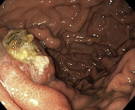
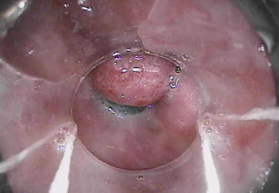
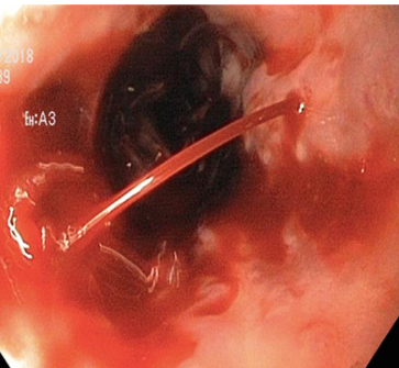
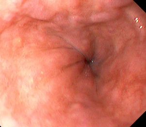
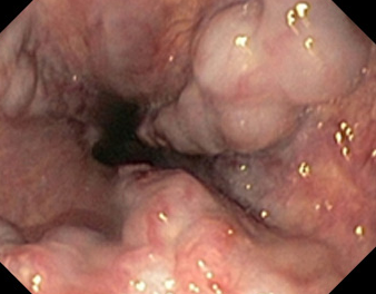
Endoscopic image after performance of splenectomy. Complete disappearance of the varices of the fundus of the stomach (Courtesy Dr. V. Penopoulos)
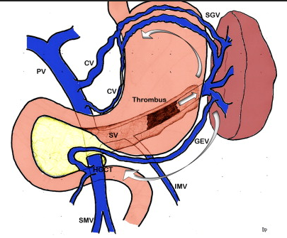
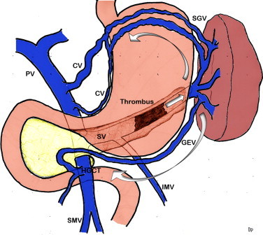
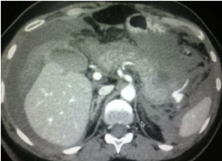
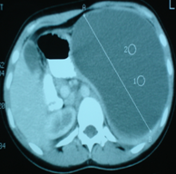
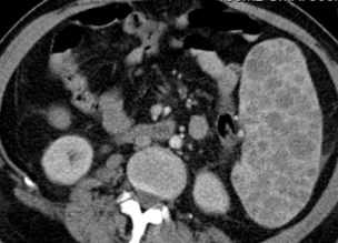
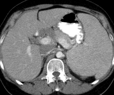
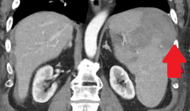
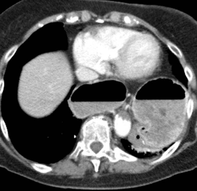
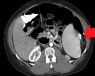
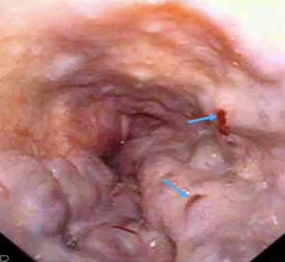
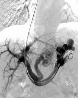
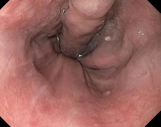
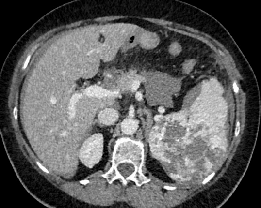
Abdominal CT scan. Visible presence of varices at the hilum of the spleen (Courtesy Dr. V. Penopoulos)
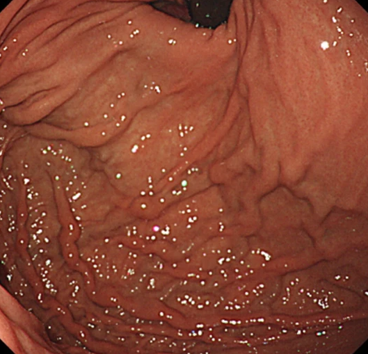
Endoscopic image after performance of splenectomy. Complete disappearance of the varices of the fundus of the stomach (Courtesy Dr. V. Penopoulos)
Endoscopic image after performance of splenectomy. Complete disappearance of the varices of the fundus of the stomach (Courtesy Dr. V. Penopoulos)
Endoscopic image after performance of splenectomy. Complete disappearance of the varices of the fundus of the stomach (Courtesy Dr. V. Penopoulos)